A black male infant was born at 40 1/7 weeks estimated gestational age via primary cesarean delivery because of failure to progress. The Apgar score was 9 at one and five minutes. A mass was noted on his right hand.
During the prenatal period, his mother was treated with valacyclovir (Valtrex) for herpes simplex virus infection. Intrauterine ultrasonography showed left kidney pyelectasis. The newborn's older sibling had a similar finding at birth.
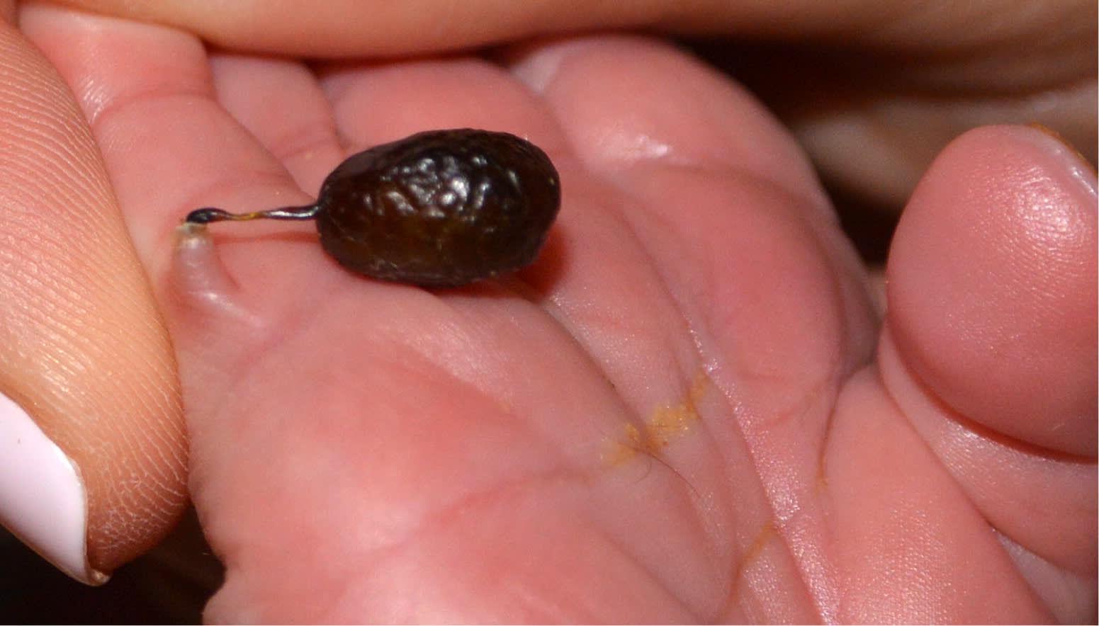
Physical examination of the newborn revealed a small, pedunculated mass that was attached by a fine stalk to the ulnar aspect of his right fifth digit (Figure 1). The mass was initially green but turned black at two hours of life. The remainder of the physical examination was unremarkable, with no other cutaneous growths or malformations noted.
Figure 1
Question
Based on the patient's history and physical examination findings, which one of the following is the most likely diagnosis?
A. Accessory (supernumerary) digit.
B. Acquired digital fibrokeratoma.
C. Infantile digital fibroma.
D. Periungual fibroma (Koenen tumor).
Discussion
The answer is A: accessory (supernumerary) digit. Rudimentary accessory digits, also known as polydactyly, typically present as small, flesh-colored papules or nodules attached by a stalk, most often on the ulnar aspect of the fifth digit (postaxial). They can also arise from the radial aspect of the thumb (preaxial).1 Accessory digits occur in approximately one out of 1,000 live births in whites, and in three to 10 out of 1,000 live births in blacks.1 They may occur as an isolated finding, with a familial pattern suggesting autosomal dominant inheritance, or as part of a syndrome with other developmental abnormalities.1,2
Complications of accessory digits include infection and necrosis. Accessory digits may appear blue with a cystic-like structure due to lymphatic obstruction.3 In this patient, the black color change may have been due to torsion or strangulation resulting in necrosis.
Accessory digits can be distinguished from other common pediatric acral masses by their presentation at birth and histologic evaluation, which reveals abundant nerve bundles in the dermis. These nerve bundles are thought to represent a small amputation neuroma that results from intrauterine amputation of the digit.2 In some cases, the lesion may also contain bone, cartilage, or a nail apparatus.2 Surgical excision is the treatment of choice.1
Acquired digital fibrokeratomas are rare, benign, solitary, firm, slow-growing nodules commonly found on the fingers and toes. They are often mistaken for other common conditions such as an accessory digit, verruca vulgaris, or cutaneous horn.4 Histology differentiates acquired digital fibrokeratomas because they contain densely packed collagen bundles often perpendicular to the skin surface (oriented vertically in the dermis).5 Acquired digital fibrokeratomas do not spontaneously resolve and require excision.4
Infantile digital fibromas are small, benign, dermal or subcutaneous nodules that develop on the dorsal or lateral aspect of the phalanges, but characteristically spare the thumb and great toe.6 They typically develop sporadically during the first year of life, although 30% of cases may be congenital.7 Histologically, they contain spindle cells with characteristic red inclusion bodies, which show up as positive on special stains for actin and vimentin.5 They often spontaneously resolve within two to three years; therefore, excision is not recommended.5
Periungual fibromas, also known as Koenen tumors, are benign, red to flesh-colored, smooth, often pedunculated skin papules or nodules that develop around the nail folds. They are more common on toenails than fingernails. The papules may enlarge, causing a visible groove distortion in the nail plate.1 Histologically, they demonstrate concentric perivascular fibrosis.5 They are considered a pathognomonic feature for tuberous sclerosis, and are part of the diagnostic criteria for the disease.8
Summary Table
| Condition | Limb abnormalities | Other characteristics |
|---|---|---|
| Accessory (super-numerary) digit | Small, flesh-colored papule or nodule attached by a stalk, most often on the ulnar aspect of the fifth digit; may have bone, cartilage, or nail apparatus | Nerve bundles |
| Acquired digital fibrokeratoma | Solitary, firm, slow-growing nodule; commonly found on the fingers and toes | Hyperkeratosis, acanthosis, hypergranulosis, spindle cells and collagen often oriented perpendicular to the skin surface |
| Infantile digital fibroma | Small dermal or subcutaneous nodule that develops on the dorsal or lateral aspect of the phalanges; the thumbs and great toes are often spared | Spindle cells with characteristic red inclusion bodies, which show up as positive on special stains for actin and vimentin |
| Periungual fibroma (Koenen tumor) | Red to flesh-colored, smooth, often pedunculated papules or nodules that develop around the nail folds; more common on toenails than fingernails | Concentric perivascular fibrosis |
The authors thank Christina N. Lawson, MD, and Banku Jairath, MD, for their assistance.
