
Am Fam Physician. 2017;95(8):519-520
Author disclosure: No relevant financial affiliations.
A 78-year-old man who had not seen a physician in many years had pain in his feet, but no history of trauma. He wore sneakers and was able to walk well without a cane or walker. He had not cut his toenails for an extended period and had poor hygiene. He was independent in activities of daily living. He had no relevant medical history and was not taking medications.
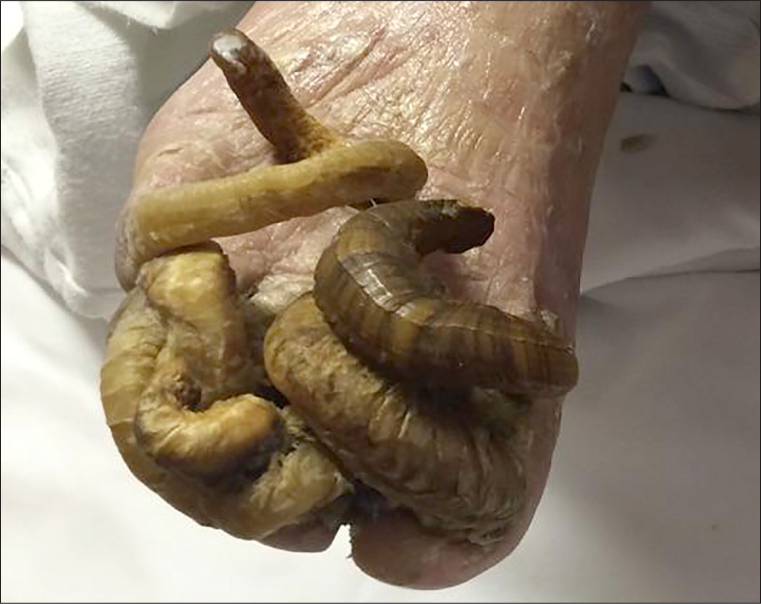
On physical examination, his lower extremities were cool with thin, dry, and scaly skin. Bilateral thick, elongated, and curved toenails 5 to 8 cm long were digging into the skin (Figure 1). Keratin debris was noted over the toes and interdigital spaces, and there was trace pitting edema on both legs. Dorsalis pedis pulses were diminished bilaterally. Sensation was grossly decreased to light touch.
Question
Discussion
The answer is B: onychogryphosis. The condition is characterized by thickened, discolored, severely elongated, curved nails with claw-like deformity (ram's horn nail). Onychogryphosis occurs mostly in older or homeless persons as a result of chronic neglect, trauma, or poor nail care.1–4 Although it occurs most commonly in the great toenails, any nail can be affected. Underlying peripheral vascular disease, poor circulation, and associated onychomycosis may occur, as in this patient.1–4
Treatment of patients with onychogryphosis includes trimming the nails with an electric burr and filing subungual hyperkeratosis. Rough edges of the toenail should be smoothed down. Other treatments include avulsion of the nail plate and surgical or chemical destruction of the nail matrix with phenol or a carbon-dioxide laser.5–7 The choice of treatment should take into account peripheral vascular circulation, treatment compliance, and potential complications. Periodic repeated treatments are usually required for maintenance.5 Any coexisting fungal infection should be treated appropriately.1,2
| Condition | Limb abnormalities |
|---|---|
| Onychauxis | Thickening of the nail plate with discoloration in older persons; may be associated with subungual hyperkeratosis of the nail bed |
| Onychogryphosis | Thickened, discolored, severely elongated nails; curled, claw-like deformity (ram's horn); underlying peripheral vascular disease, poor circulation, or associated onychomycosis may occur |
| Onychorrhexis | Increased nail brittleness with longitudinal ridging and fissuring of the nail plate |
| Onychoschizia | Splitting of the distal lamella of the nail horizontally at the free edge; often due to water or detergent exposure |
| Trachyonychia | Thinned nail with longitudinal ridges, pitting, and roughening of the proximal nail surface |
Onychoschizia is splitting of the distal lamella of the nail horizontally at the free edge. It is often due to frequent water or detergent exposure.4
