
Am Fam Physician. 2017;95(10):667-668
Author disclosure: No relevant financial affiliations.
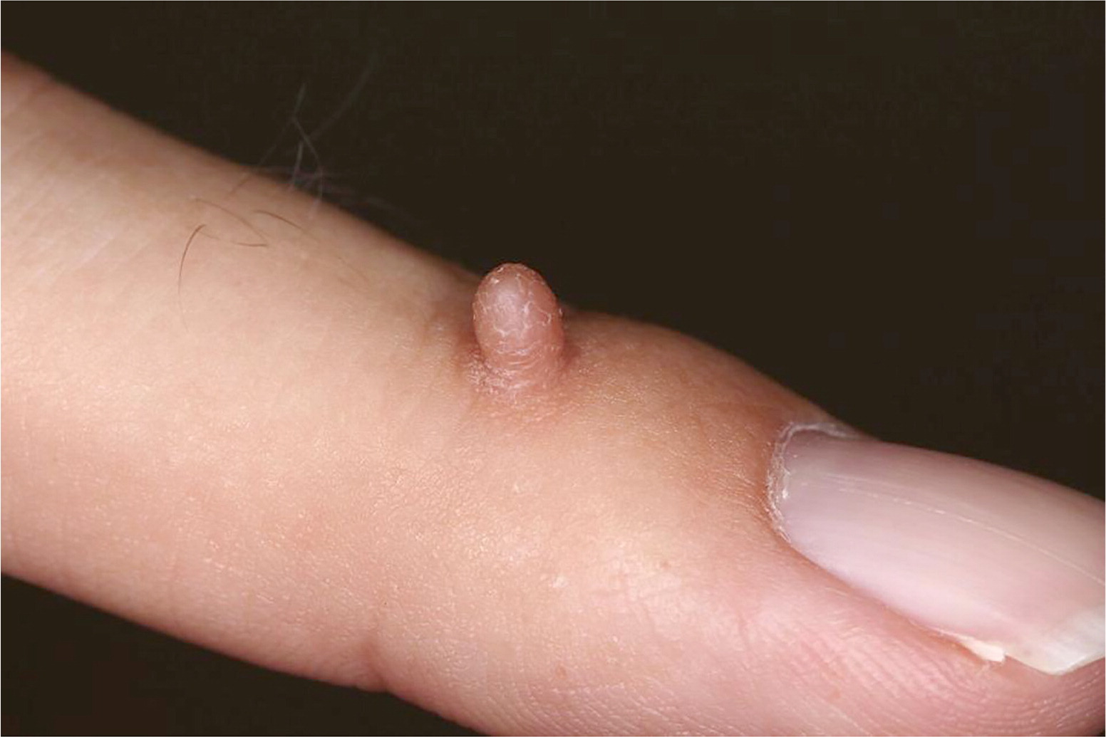
A 36-year-old man presented with a self-described “wart” on his finger. It had been present since childhood, although he did not know if he was born with it. The lesion was a nuisance, but it did not itch or bleed and was nontender. It did not resolve with cryotherapy performed approximately 20 years earlier. He had no other similar lesions or significant medical history, including skin disease.
Physical examination revealed a 3-mm, exophytic, firm, flesh-colored filiform papule on the dorsum of the left third finger with slight scaling (Figure 1).
Question
Discussion
The answer is A: acquired digital fibrokeratoma. Acquired digital fibrokeratomas are uncommon benign pedunculated neoplasms. They are usually 3 to 5 mm in diameter, but lesions larger than 3 cm have been documented.1 A collarette of raised skin encircling the base differentiates digital fibrokeratomas from similar lesions. This creates the appearance of an annular moat around the lesion.2 Acquired digital fibrokeratomas are usually found on the fingers and toes.2,3 Rarely, they involve the nail.4
There is no definitive etiology for acquired digital fibrokeratomas, but they are thought to be associated with subclinical trauma.2 The incidence is unknown. Although they seem to occur most often in middle-aged adults, there are documented cases in patients 12 to 70 years of age.2 Histologically, a hyperkeratotic stratum corneum is seen overlying a dome-shaped papule; acanthosis may be present.2 Acquired digital fibrokeratomas do not resolve spontaneously, and complete excision is the treatment of choice.5
Common warts (verruca vulgaris) are the most common neoplasm of the hand and are associated with human papillomavirus.5 They present as flesh-colored, irregular papules that may be sessile or exophytic. They have characteristic verrucous surface changes. Common warts are less hyperkeratotic than cutaneous horns.
Cutaneous horns are filiform projections of cornified epidermis that may possess various histologic lesions at their base. They can be found anywhere on the body but favor sun-exposed areas. These hyperkeratotic, exophytic, hard papules are usually small but can grow larger than 10 cm.2
Myxoid cysts (digital mucous cysts) appear as a solitary, flesh-colored papule or nodule over the distal interphalangeal joint. They may develop over the proximal nail fold, causing grooves in the nail plate. The cyst is filled with clear, viscous fluid.
Supernumerary digits (polydactyly) range in appearance from well-developed recognizable fingers to rudimentary pedunculated, soft, flesh-colored papules. They may be difficult to distinguish from digital fibrokeratomas when they are small and poorly developed; biopsy may be required.
| Lesion | Gross appearance | Location |
|---|---|---|
| Acquired digital fibrokeratoma | Collarette of raised skin encircling the base, creating the appearance of an annular moat around the lesion | Fingers and toes, areas of subclinical trauma |
| Common wart (verruca vulgaris) | Flesh-colored, verrucous papule; associated with human papillomavirus | Most common on hands but can occur anywhere on the body |
| Cutaneous horn | Exophytic, filiform, cornified papule; very hard | Sun-exposed areas |
| Myxoid cyst (digital mucous cyst) | Solitary, flesh-colored, fluid-filled papule or nodule | Over distal interphalangeal joint or proximal nail fold |
| Supernumerary digit (polydactyly) | Rudimentary form appears as a pedunculated, soft, flesh-colored exophytic papule | Hands and feet |
