A 33-year-old woman presented with a growth on her eye that had been present for several years. She believed that it had grown over the previous six to eight months. She was asymptomatic other than seasonal pruritus, especially during the summer. There was no localized pain, discharge, or vision changes.
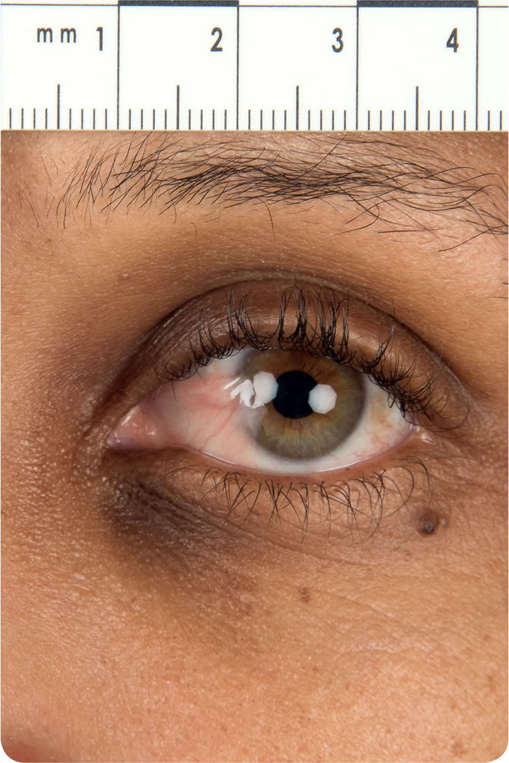
Physical examination revealed a small, clear corneal growth on the left eye that extended medially toward the pupil (Figure 1). There were no abnormal findings on neurologic examination.
FIGURE 1
Question
Based on the patient's history and physical examination findings, which one of the following is the most likely diagnosis?
A. Conjunctival neoplastic lesion.
B. Conjunctivitis.
C. Episcleritis.
D. Pinguecula.
E. Pterygium.
Discussion
The answer is E: pterygium. The pterygium is a triangular wedge of soft, nearly flat fibrovascular conjunctival tissue that starts medially on the nasal conjunctiva and extends laterally onto the cornea.1 It is white and amorphous or thick. It may have redness or irritation. The lesion usually is not noticed unless it appears white against a colored iris or red because of a vascular component. It is typically bilateral but may be unilateral.
Pterygium is usually asymptomatic but may cause mild visual impairment. It can extend onto the cornea and impair vision through induced astigmatism. If the lesion is larger than 3.5 mm (greater than halfway to the center of the pupil in a typical cornea, 11 to 12 mm), it will cause blurriness. Pterygium can restrict eye movement if the inflammation causes the conjunctiva and overlying eyelid to stick together. Pterygium may be caused by chronic exposure to ultraviolet light.2 The prevalence of pterygium varies from 1% to 25%.3 Treatment depends on the size. A small pterygium may be treated symptomatically with artificial tears. A large pterygium may require surgical excision if it is associated with visual impairment.3
A conjunctival neoplastic lesion may present similarly to a pterygium.4 A neoplastic lesion is more vascular and irregular in consistency and shape, and is typically unilateral. It usually arises from the interpalpebral fissure.
Conjunctivitis is an inflammation of the bulbar and/or palpebral conjunctiva. It can be infectious (viral or bacterial) or allergic. Infectious conjunctivitis presents as a red eye with mucoid or serous discharge and a burning, sandy, gritty sensation. It is usually unilateral and does not impair vision. However, allergic conjunctivitis is typically bilateral and presents as red eyes, watery discharge, and itching.
Episcleritis is an inflammation of the episcleral tissue between the conjunctiva and sclera. It is characterized by the abrupt onset of redness, irritation, and eye watering. It is associated with normal vision and is typically not painful. Patients with suspected episcleritis should be referred to an ophthalmologist.
A pinguecula is conjunctival degeneration presenting as a yellowish, slightly raised conjunctival lesion arising at the limbal conjunctiva. It is differentiated from a pterygium in that it arises from the limbus and remains confined to the conjunctiva without corneal involvement.5 There is a space between the pinguecula and the edge of the cornea.6
SUMMARY TABLE
| Condition | Characteristics |
|---|---|
| Conjunctival neoplastic lesion | More vascular and irregular in consistency and shape than a pterygium; typically unilateral; usually arises from the interpalpebral fissure |
| Conjunctivitis | Inflammation of the bulbar and/or palpebral conjunctiva; red eye with mucoid or serous discharge; burning, sandy, gritty sensation; no associated visual impairment; etiology may be viral, bacterial, or allergic |
| Episcleritis | Inflammation of the episcleral tissue between the conjunctiva and sclera; abrupt onset of redness, irritation, and eye watering |
| Pinguecula | Conjunctival degeneration presenting as a yellowish, slightly raised conjunctival lesion arising at the limbal conjunctiva |
| Pterygium | Triangular wedge of soft, nearly flat fibrovascular conjunctival tissue that starts medially on the nasal conjunctiva and extends laterally onto the cornea; it is white and amorphous or thick; may have redness or irritation |
The views expressed in this article are those of the authors and do not necessarily reflect the official policy or position of the Department of the Navy, Department of Defense, or the U.S. government.
We are military services members. This work was prepared as part of our official duties. Title 17 U.S.C. 105 provides that copyright protection under this title is not available for any work of the U.S. government.
