Clinical Question
Does the use of dermoscopy aid in the detection of melanoma in adults?
Evidence-Based Answer
The addition of dermoscopy to in-person visual inspection of skin lesions increases specificity and sensitivity in the detection of melanoma. In-person evaluation with dermoscopy is more accurate than image-based assessment. A published algorithm meant to assist in dermoscopic interpretation does not improve the accuracy of diagnosis, whereas dermoscopic training does increase diagnostic accuracy. There is currently insufficient evidence to assess the accuracy of dermoscopy in the primary care setting.1 (Strength of Recommendation: A, based on consistent, good-quality patient-oriented evidence.)
Practice Pointers
Dermoscopy (also known as dermatoscopy or epiluminescence microscopy) describes the use of a noninvasive handheld magnification instrument to evaluate skin lesions. Dermoscopy reveals colors and microstructures not visible to the naked eye that correspond to histologic attributes.2 Dermoscopic findings may prompt biopsy of suspicious lesions or provide reassurance for benign lesions. Images from dermoscopy can be evaluated in the presence of the patient (i.e., dermoscopic in-person evaluation) or saved and reviewed at a later time (i.e., dermoscopic image-based evaluation). The term “visual inspection” describes routine examination performed without the aid of a dermatoscope or other device. The Surveillance, Epidemiology, and End Results Program estimated 96,480 new cases of melanoma in the United States in 2019, accounting for 5.5% of all new cancer cases, making melanoma the fifth most frequent new cancer diagnosis.3
This Cochrane review included 104 studies and 42,788 total skin lesions suspected to be melanoma.1 The percentage of patients with melanoma ranged between 1% and 41% for dermoscopic in-person studies (median = 12%) and between 3% and 61% in studies using dermoscopic images (median = 24%). The diagnosis of melanoma was made by histology, and the absence of melanoma was confirmed by histology or by follow-up over time to ensure the skin lesion remained negative for melanoma. In four studies, the absence of melanoma was confirmed by expert diagnosis. Almost all of the studies were carried out in specialty offices, with only four of the studies conducted in a primary care setting.
The main results were based on 83 publications that provided 86 data sets, including accuracy data for the detection of invasive melanoma and atypical intraepidermal melanocytic variants. Twenty-six studies provided information on the accuracy of dermoscopy added to in-person visual inspection, and 60 studies provided information on dermoscopic image-based evaluation. The accuracy of diagnosis using in-person visual inspection with dermoscopy was significantly higher than evaluation of dermoscopic images, with a diagnostic odds ratio for in-person dermoscopy more than four times that of the image-based diagnosis (relative diagnostic odds ratio = 4.6; 95% CI, 2.4 to 9.0; P < .001). The review analyzed in-person and image-based studies separately because of this difference.
The reviewers also found no apparent benefit for accuracy with the use of an algorithm (e.g., “ABCD” or pattern analysis) to assist in dermoscopic interpretation for in-person or image-based evaluations. General practitioners demonstrated decreased accuracy compared with specialists in two in-person studies and three image-based studies. Regardless of the different types and length of dermoscopic training, all of the six training interventions included in the review resulted in increased sensitivity of diagnosis, with little effect on specificity in five of the six studies.
Dermoscopy is a useful tool to support visual inspection of suspicious skin lesions in specialty care offices with a trained practitioner. Further evaluation in the primary care setting and determination of the optimal approach to training are needed. Currently no accepted guidelines promote the use of dermoscopy in primary care in the United Kingdom, United States, or Canada. The 2015 Suspected Cancer: Recognition and Referral guideline from the National Institute for Health and Care Excellence recommends that patients be referred for biopsy “using a suspected cancer pathway referral (for an appointment within two weeks) if dermoscopy suggests melanoma of the skin.” 4
SUMMARY TABLE In-Person Visual Inspection Alone vs. Visual Inspection Plus Dermoscopy for the Detection of Melanoma in Adults
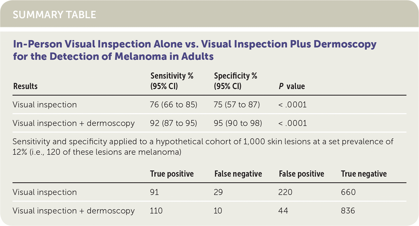
| Results | Sensitivity % (95% CI) | Specificity % (95% CI) | P value | |
| Visual inspection | 76 (66 to 85) | 75 (57 to 87) | < .0001 | |
| Visual inspection + dermoscopy | 92 (87 to 95) | 95 (90 to 98) | < .0001 | |
| Sensitivity and specificity applied to a hypothetical cohort of 1,000 skin lesions at a set prevalence of 12% (i.e., 120 of these lesions are melanoma) | ||||
| True positive | False negative | False positive | True negative | |
| Visual inspection | 91 | 29 | 220 | 660 |
| Visual inspection + dermoscopy | 110 | 10 | 44 | 836 |
The practice recommendations in this activity are available at http://www.cochrane.org/CD011902.
