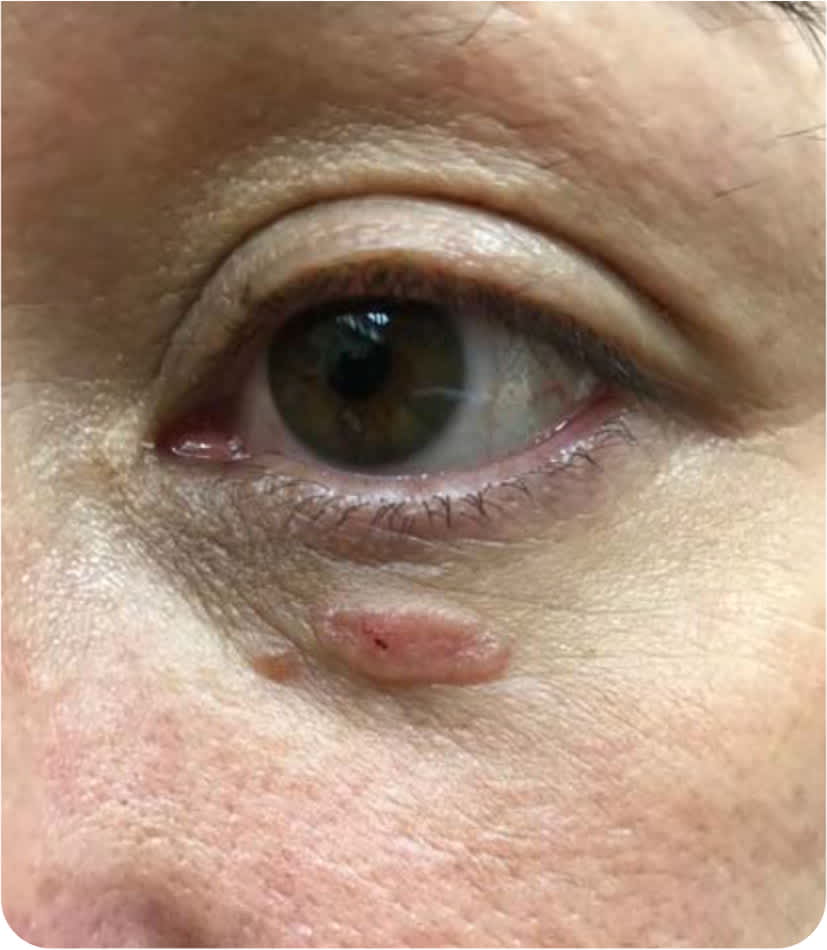
A 46-year-old woman presented with a mass under her eye that had been growing slowly for approximately three years. It was pruritic but not associated with pain or bleeding. She had no significant medical history aside from hyperlipidemia.
Physical examination revealed a pink papule on her left lower eyelid, which had a central punctation with rolled borders (Figure 1).
FIGURE 1
Question
Based on the patient's history and physical examination findings, which one of the following is the most likely diagnosis?
A. Hidrocystoma.
B. Lipoid proteinosis.
C. Nodular basal cell carcinoma.
D. Syringoma.
E. Xanthelasma.
Discussion
Answer is C: nodular basal cell carcinoma. Basal cell carcinoma is the most common cancer worldwide.1 Lifetime risk in the United States is approximately 20%, and the annual incidence is estimated at 8%.1 Risk factors include Fitzpatrick skin types I and II. The predominant environmental risk factor is exposure to ultraviolet radiation.2 Subtypes include nodular, superficial, infundibulocystic, fibroepithelial, morpheaform, and infiltrative.2 Nodular basal cell carcinoma is the most common subtype, accounting for 50% to 80% of basal cell carcinoma lesions.1
Nodular basal cell carcinoma presents as a translucent, pink, or pearly well-circumscribed papule or nodule with rolled borders. Lesions are often associated with telangiectasias. Lesions may be asymptomatic, or they may ulcerate, bleed, or be pruritic. The face (i.e., cheeks, nasolabial folds, forehead, and eyelids) is the most common site for basal cell carcinoma; however, it may occur on any hair-bearing surface.3
Skin biopsy is the diagnostic standard for nodular basal cell carcinoma. The potential for metastasis is low, and treatment is through local control. Excision with 4-mm margins is effective for most primary tumors occurring on the neck, trunk, and extremities. Mohs surgery is the preferred treatment for cosmetically sensitive skin cancers; surgically challenging areas such as the face, pretibial region, and dorsal hands; recurrent basal cell carcinomas; and high-risk tumors. High-risk tumors are classified according to size, histologic subtype, border definition, and immunosuppression.3 Appropriate use criteria have been created to determine if a basal cell carcinoma is suitable for Mohs surgery.4 These criteria are available at https://www.nccn.org/professionals/physician_gls/pdf/nmsc.pdf. Curettage and electrodesiccation, topical chemotherapy, cryosurgery, and photodynamic therapy can also be used to treat low-risk tumors.3 The National Comprehensive Cancer Network guidelines can direct management.5
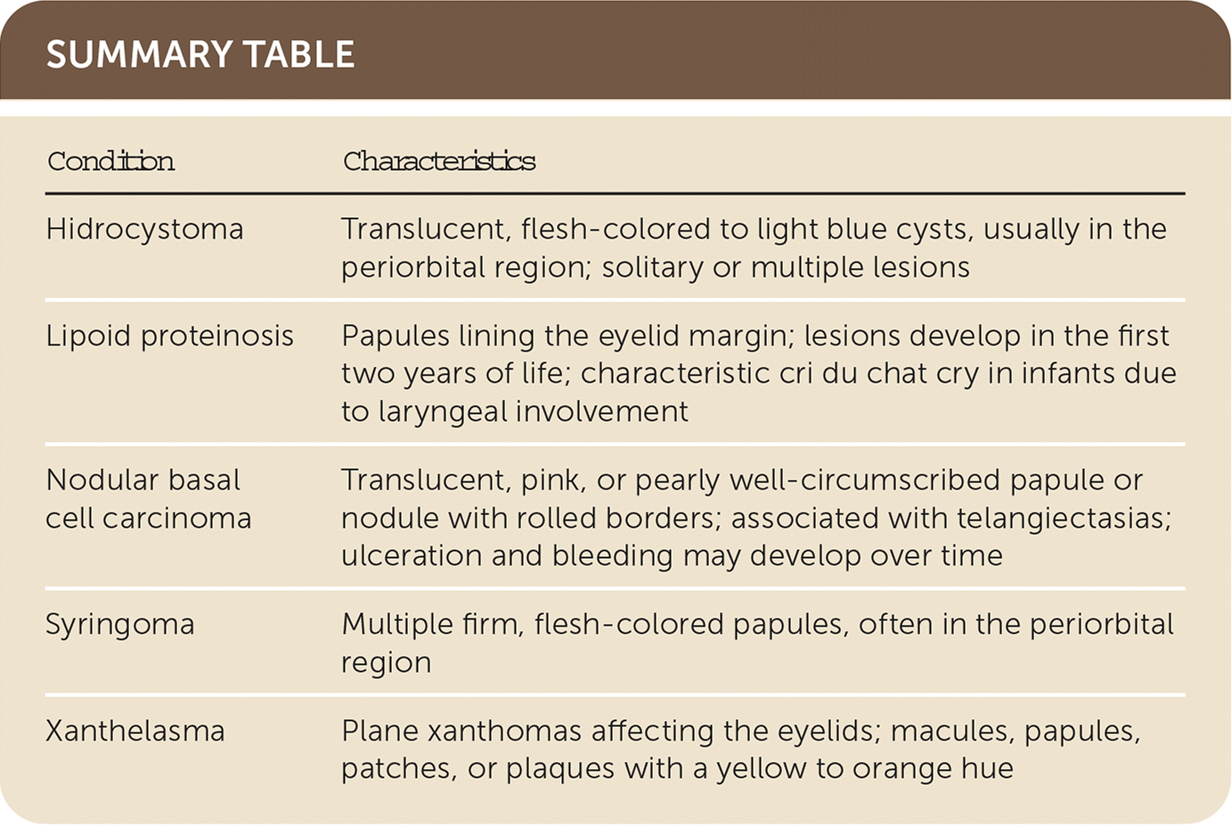
Hidrocystomas are benign cystic tumors arising from apocrine or eccrine glands. They typically present in the periorbital region as translucent, flesh-colored to light blue cysts. Apocrine hidrocystomas usually present as solitary lesions, whereas eccrine hidrocystomas can present as solitary or multiple lesions.3
Lipoid proteinosis is an autosomal recessive deposition disorder that results in hyaline deposits in multiple organs, including the skin, oral mucosa, larynx, and brain. Cutaneous lesions develop within the first two years of life. Classic findings include papules lining the eyelid margin and a characteristic cri du chat (i.e., a high-pitched, cat-like cry due to laryngeal involvement) in infants.3
Syringomas are benign adnexal neoplasms that present as multiple firm, flesh-colored papules, often in the periorbital region, particularly the eyelids.3
Xanthelasma is characterized by plane xanthomas affecting the eyelids. The lesions can present as macules, papules, patches, or plaques with a yellow to orange hue. Xanthelasma is associated with hyperlipidemia in about one-half of patients.3
SUMMARY TABLE
| Condition | Characteristics |
|---|---|
| Hidrocystoma | Translucent, flesh-colored to light blue cysts, usually in the periorbital region; solitary or multiple lesions |
| Lipoid proteinosis | Papules lining the eyelid margin; lesions develop in the first two years of life; characteristic cri du chat cry in infants due to laryngeal involvement |
| Nodular basal cell carcinoma | Translucent, pink, or pearly well-circumscribed papule or nodule with rolled borders; associated with telangiectasias; ulceration and bleeding may develop over time |
| Syringoma | Multiple firm, flesh-colored papules, often in the periorbital region |
| Xanthelasma | Plane xanthomas affecting the eyelids; macules, papules, patches, or plaques with a yellow to orange hue |
The opinions expressed belong solely to the authors. They should neither be interpreted as representative of nor endorsed by the Uniformed Services University of the Health Sciences, the U.S. Navy, the Department of Defense, or any other federal government agency.
