
Am Fam Physician. 2020;101(11):689-690
Author disclosure: No relevant financial affiliations.
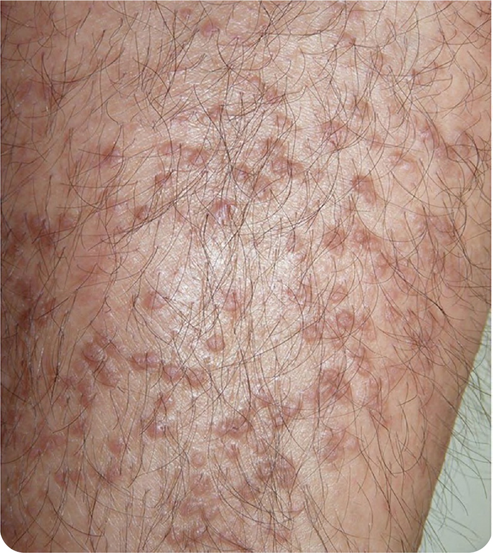
A 36-year-old man presented with an intensely pruritic rash on both lower legs that had been present for three years. He had a history of asthma and moderate atopic dermatitis since childhood. The rash coincided with progressive worsening of his atopic dermatitis. It was associated with generalized pruritus that caused him to scratch, especially on his lower legs where the eruption was most intense. Physical examination revealed multiple hyperkeratotic papules on the lower limbs, particularly the pretibial areas (Figure 1).
Question
Discussion
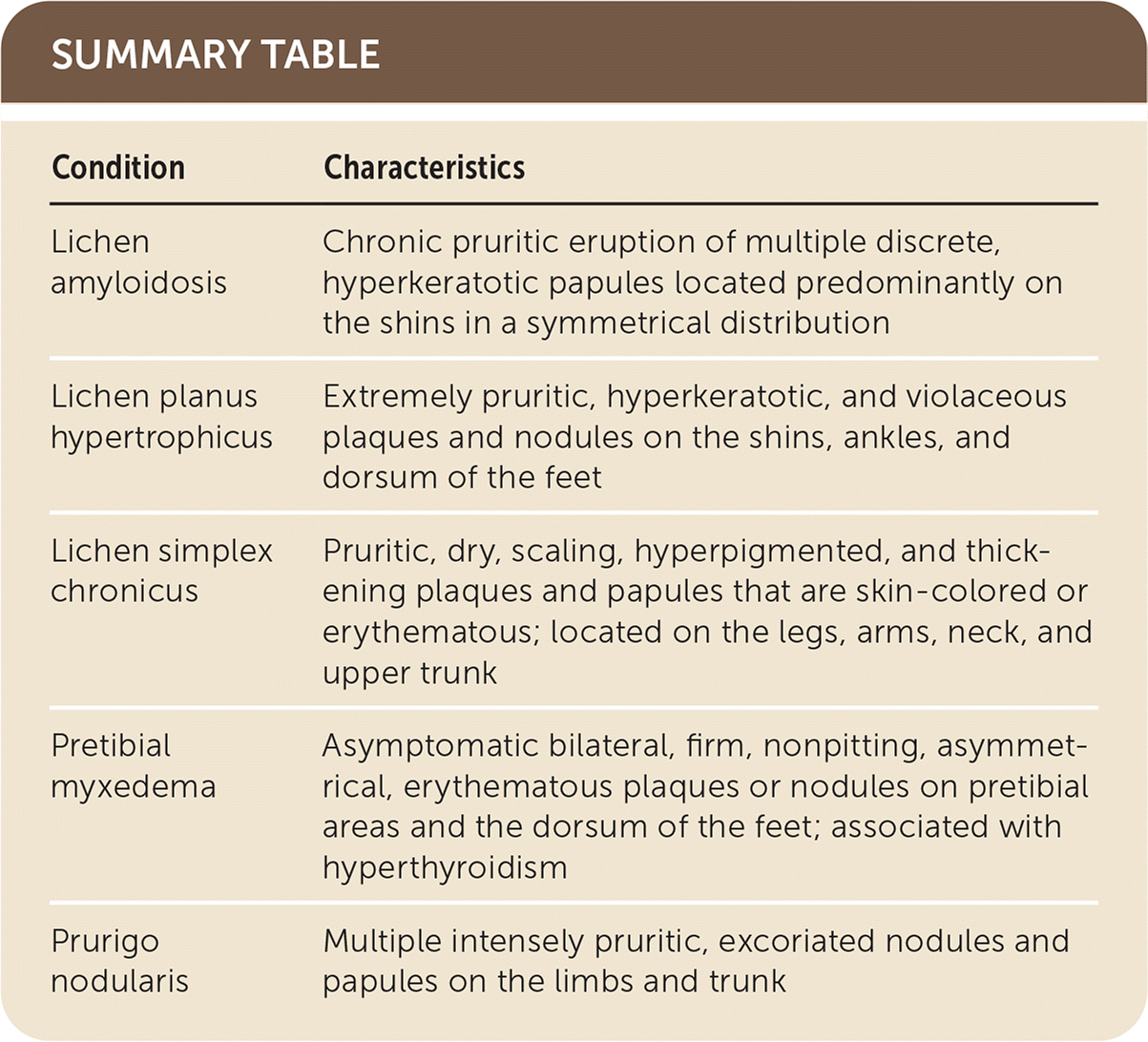
The answer is A: lichen amyloidosis. Primary localized cutaneous amyloidosis is a chronic pruritic skin disorder characterized by amyloid deposition in the dermis without systemic involvement. It is most prevalent in Southeast Asia and South America.1,2 Lichen amyloidosis is a subtype that mainly affects middle-aged adults, especially men with darker skin. It is characterized by a chronic pruritic eruption of multiple discrete, hyperkeratotic papules located predominantly on the shins in a symmetrical distribution.
Although the diagnosis of lichen amyloidosis is usually made clinically, histopathology characteristically shows deposits of hyaline material in the papillary dermis corresponding to amyloid, with positive results on Congo red testing. Most cases are idiopathic, but lichen amyloidosis may be associated with multiple endocrine neoplasia type 2A, atopic dermatitis, and autoimmune diseases.1–3 This patient's laboratory findings were within normal limits (blood cell count, biochemistry, thyroid-stimulating hormone, HIV), except for an immunoglobulin E level of 2,575 IU per mL (normal = 100 IU per mL or less). The pathogenesis of lichen amyloidosis is not clear, but the amyloid deposits are from degenerated keratin peptides of apoptotic keratinocytes. Prolonged epidermal trauma, including scratching, appears to be the most important factor in the pathogenesis.3
Current therapies for lichen amyloidosis have high relapse rates. Treatment is directed at relieving the pruritus and addressing the underlying atopic dermatitis. Narrow band ultraviolet B phototherapy appears to be the best treatment option. Other treatments include topical and oral corticosteroids, topical calcineurin inhibitors, antihistamines, cyclosporine (Sandimmune), and fractional carbon-dioxide laser therapy.3
Lichen planus hypertrophicus classically involves the shins, ankles, and dorsum of the feet. It is characterized by extremely pruritic, hyperkeratotic, and violaceous plaques and nodules in a symmetrical distribution. The condition may be associated with chronic venous disease.
Lichen simplex chronicus is a common dermatologic disorder secondary to constant scratching. It is often associated with atopic dermatitis and predominantly affects women 30 to 50 years of age. It manifests as pruritic, dry, scaling, hyperpigmented, and thickening plaques and papules that are skin-colored or erythematous. It involves sites that are easily reached for scratching, most commonly the legs, arms, neck, and upper trunk.
Pretibial myxedema is generally associated with autoimmune thyroid disorders, mainly hyperthyroidism. It is more common in women and involves the pretibial areas and the dorsum of the feet. It is usually asymptomatic. Early lesions are bilateral, firm, nonpitting, asymmetrical plaques or nodules that are skin-colored or erythematous. The lesions may coalesce to form scaly, thickened, and hardened areas of the skin.
Prurigo nodularis presents as multiple intensely pruritic, excoriated nodules and papules on the limbs and trunk in the setting of generalized chronic pruritus. It can appear at any age but usually occurs between 20 and 60 years. It is most common in women.4
| Condition | Characteristics |
|---|---|
| Lichen amyloidosis | Chronic pruritic eruption of multiple discrete, hyperkeratotic papules located predominantly on the shins in a symmetrical distribution |
| Lichen planus hypertrophicus | Extremely pruritic, hyperkeratotic, and violaceous plaques and nodules on the shins, ankles, and dorsum of the feet |
| Lichen simplex chronicus | Pruritic, dry, scaling, hyperpigmented, and thickening plaques and papules that are skin-colored or erythematous; located on the legs, arms, neck, and upper trunk |
| Pretibial myxedema | Asymptomatic bilateral, firm, nonpitting, asymmetrical, erythematous plaques or nodules on pretibial areas and the dorsum of the feet; associated with hyperthyroidism |
| Prurigo nodularis | Multiple intensely pruritic, excoriated nodules and papules on the limbs and trunk |
