
Am Fam Physician. 2021;104(2):199-200
Author disclosure: No relevant financial affiliations.
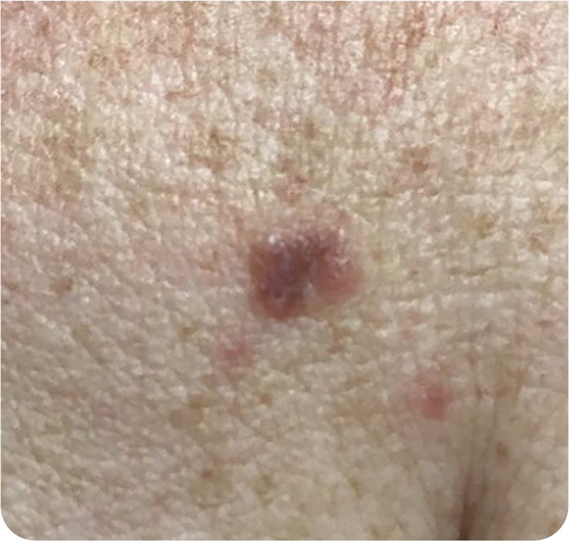
A 54-year-old patient presented with a new lesion on the upper chest that had been present for three months. The lesion was solitary and slightly pruritic.
The patient had no trauma to the involved area and no family history of melanoma. The patient had a history of skin lesions that were biopsied but determined to be benign. The patient reported multiple severe sunburns over several years and had spent numerous summers playing outside as a child. The patient used sunscreen regularly.
Physical examination revealed a red, slightly raised papule on the anterior chest wall, near the left breast (Figure 1).
Question
Based on the patient's history and physical examination findings, which one of the following is the most likely diagnosis?
A. Actinic keratosis.
B. Basal cell carcinoma.
C. Bowen disease.
D. Lichenoid keratosis.
E. Seborrheic keratosis.
Discussion
The answer is D: lichenoid keratosis. Lichenoid keratosis normally occurs in middle-aged and older patients and is more common in those with frequent sun exposure and sun-damaged skin. Lichenoid keratosis typically presents as a solitary asymptomatic lesion on the skin, ranging from 5 to 20 mm in diameter.1 It is a slightly hardened, plaque-like lesion that is normally erythematous but can be brown or sometimes violaceous.1
Because of the wide-ranging clinical appearance, lichenoid keratosis is often misdiagnosed.1 The condition is benign and does not have the histologic characteristics of basal cell carcinoma.2 Because the presentation of lichenoid keratosis is similar to that of basal cell carcinoma, a biopsy may be required to confirm the correct diagnosis. If desired or for cosmetic reasons, lichenoid keratosis can be removed through cryotherapy or excision.
Actinic keratosis is a distinct, hyperkeratotic lesion on the skin due to confined proliferation of keratinocytes at the dermoepidermal junction. This proliferation causes a disturbance in the differentiation of the epidermis, which commonly presents as a rough, scaly patch of skin.3
Basal cell carcinoma generally presents on sun-exposed skin, most notably on the head and neck. It presents as a pearly papule or nodule with overlying telangiectasia and a translucent rolled border.4
Bowen disease, the in situ form of squamous cell carcinoma, presents as a nonpigmented, slowly growing, erythematous, well-demarcated plaque with a scaly or crusty surface that may be eroded or ulcerated.5 Bowen disease is common on the lower extremities of older patients with sun-damaged skin. It is more common in women.5
Seborrheic keratoses are a specific type of benign cutaneous tumors, with varying clinical and histopathologic presentations. They develop from the proliferation of epidermal keratinocytes and are common in people 40 years and older.6
| Condition | Characteristics |
|---|---|
| Actinic keratosis | Distinct, rough, scaly, hyperkeratotic lesion |
| Basal cell carcinoma | Pearly papule or nodule with overlying telangiectasia and a translucent rolled border; most common on the head and neck |
| Bowen disease | Nonpigmented, slowly growing, erythematous, well-demarcated plaque with a scaly or crusty surface that may be eroded or ulcerated; common on the lower extremities of older patients with sun-damaged skin |
| Lichenoid keratosis | Solitary asymptomatic lesion; 5 to 20 mm in diameter; slightly hardened, plaque-like lesion; erythematous to brown or sometimes violaceous |
| Seborrheic keratosis | Benign cutaneous tumors that develop from the proliferation of epidermal keratinocytes; clinical presentation varies |
