
Am Fam Physician. 1998;58(8):1829-1830
Breathing Space
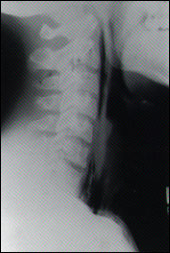
An 18-year-old man with a remote history of asthma presented to our office with the acute onset of dyspnea and retrosternal chest pain. The patient had been evaluated previously by another physician and treated with nebulized albuterol for a presumed asthma exacerbation. Later that day, he presented to our emergency department with persistent symptoms. He had no history of trauma and no symptoms of fever, cough or prior respiratory infection. Vital signs were: temperature, 36.4°C (97.6°F); blood pressure, 126/66 mm Hg; pulse, 86; respiration, 17. Pulse oximetry measured 100 percent oxygen saturation on room air. The physical examination was remarkable for subcutaneous emphysema extending to his mandible bilaterally, and Hamman's sign. Radiographs of the patient's chest and neck are shown in the accompanying figures.
Question
Discussion
The answer is A: spontaneous pneumomediastinum. Spontaneous pneumomediastinum is a rare, infrequently reported syndrome. It was first described by Hamman in 1939.1 It results most commonly from the rupture of pulmonary alveoli, which allows air to dissect along connective tissue planes into the mediastinum. This may also occur as a complication of mechanical ventilation. Other causes include blunt trauma to the ribs or vertebrae, or traumatic rupture of the esophagus or the tracheobronchial tree.2
Predisposing factors for the development of a spontaneous pneumomediastinum include obstructive lung disease, physical exertion and respiratory infection.3,4 The most frequent symptoms are retrosternal pain, dyspnea, dysphagia, and neck pain or a feeling of fullness.3,5,6 The physical examination may reveal subcutaneous emphysema extending to the neck or throughout the body, diminished heart sounds and pulsus paradoxus.3 Hamman's sign is the crunching noise that coincides with each heartbeat; this sign is present in approximately 50 percent of affected patients.3
A parallel line of lucency along the cardiac border is seen on radiographs, representing air between the heart and the mediastinal pleura (figure, top right). The air may dissect through tissue planes to the neck as seen in the patient here (figures, left) or to distant sites, including the extremities.6 Treatment of spontaneous pneumomediastinum includes bed rest and observation if the patient is stable.7 Beta agonists and antibiotics are indicated only if there are coincidental signs of an asthma exacerbation or an infection.
Surgical management is necessary only in severe cases of spontaneous pneumomediastinum with hemodynamic compromise, or in patients with esophageal involvement, tracheal rupture or a concomitant significant pneumothorax. Tension pneumothorax would be an indication for needle decompression. Complete reabsorption of the mediastinal air is typical and usually occurs over a period of one to two weeks.3,5 Spontaneous pneumomediastinum may recur. Because no predisposing factors for recurrence are known, restriction of activity is not currently recommended.3,8