
Am Fam Physician. 1999;59(11):3153-3154
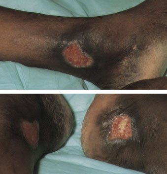
A 37-year-old man with a history of sickle cell anemia presented with chronic, non-healing ulcers (see the accompanying figures) over the lower aspects of both legs. These ulcers started spontaneously about one year before his presentation, and they were slowly enlarging. On examination, he had well-defined, superficial, non-tender ulcers measuring 4 × 2 cm near the medial malleoli of both legs. The ulcers had clean bases and increased pigmentation surrounding the bases. The patient had no varicose veins or abnormal peripheral pulses.
Question
Discussion
The answer is C: sickle cell ulcers. This patient has homozygous sickle cell anemia, presenting with sickle cell ulcers.1 Leg ulceration is a common complication in patients with homozygous sickle cell anemia. The ulcers appear either spontaneously or as a result of local trauma, and they are chronic. Possible contributing factors are obstruction of vessels by sickled cells, increased venous and capillary pressure, bacterial infection and reduced oxygen-carrying capacity of the blood.1
These ulcers are common in males, and their incidence increases significantly with advancing age. They occur more often in patients with lower hemoglobin levels and lower fetal hemoglobin levels; patients with SS and SS α-thalassemia have the highest ulcer prevalence rate of all genotypes. These ulcers occur with equal frequency over both medial and lateral aspects of the lower extremities.1 Treatment modalities include local ulcer care, antibiotics, skin grafting and transfusion therapy.1
Venous ulcers usually develop above the medial malleoli, in the area drained by incompetent perforating veins. These ulcers are well-demarcated with irregular shaggy borders and surrounding dermatitis, pigmentation and, sometimes, cellulitis.
Ischemic (arterial) ulcers occur in patients with peripheral vascular disease, usually in the distal parts of the extremities. These ulcers are painful, punched out, deep and often filled with tissue slough at the base. Associated chronic ischemic changes, such as loss of hairs, and pale and atrophic skin are usually present in the affected limb. Peripheral pulses are either diminished or absent.
Neuropathic ulcers are common in patients who have diabetes with severe neuropathy. Common sites are pressure-bearing areas such as the heels, the heads of metatarsals and the great toes. The ulcers are well-demarcated and usually surrounded by thick callus. The extremities are warm, numb and dry, with normal peripheral pulses.
Factitious ulcers are an unlikely diagnosis in this patient because there was no history to suggest this unusual and rare behavior.