A 30-year-old man with idiopathic thrombocytopenic purpura (ITP) and idiopathic constrictive pericarditis presented with an asymptomatic skin rash on his back, neck and shoulders. The rash first appeared three days earlier on his neck and had gradually progressed to both shoulders and his entire back. He did not have fever, cough, sputum or shortness of breath. As treatment for the ITP, he had been taking prednisone (50 mg daily) for two weeks before the onset of the rash. About 10 days before his presentation, he developed dermatomal herpes zoster infection over the left side of the chest and back, which was successfully treated with a standard regimen of oral acyclovir.
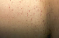
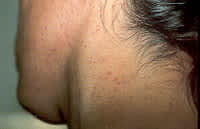
On examination, the patient was found to have multiple, discrete erythematous papules (2 to 3 mm in size) covering his entire back, shoulders and neck (see the accompanying photographs). Most of the lesions were monomorphic papules, and some were papulopustules; no vesicles were noted. He also had healed, crusted zoster rash on the left upper back and chest. His face was free of lesions. Tzanck smear and skin biopsy were performed. Tzanck smear did not show multi-nucleated giant cells. The skin biopsy result was reported to be folliculitis.
Question
Given the patient's history and the physical appearance of the rash, which one of the following would be the most likely diagnosis?
A. Disseminated herpes zoster.
B. Steroid acne.
C. Acne vulgaris.
D. Molluscum contagiosum.
E. Staphyloccocal folliculitis.
Discussion
The answer is B: steroid acne. Steroid acne is a self-limiting skin condition caused by systemic and topical corticosteroid therapy.1 It is not an uncommon problem, especially in oncologic treatment regimens and organ-transplant patients.1 In most cases, the rash appears within two weeks of the onset of corticosteroid therapy and regresses without scars when the drug is discontinued.1 Typical lesions of steroid acne are 1 to 3 mm dome-shaped papules and papulopustules, which are distributed over the face, upper trunk and upper extremities, and are monomorphic in appearance.1 Histopathology shows folliculitis. The pathogenesis of this condition is still controversial, but most experts believe that folliculitis is a common pathologic feature.1
Disseminated herpes zoster infection can be defined as more than 20 lesions outside the primary dermatomal zoster.2 It is rare in healthy persons but common in immunosuppressed patients, and is sometimes associated with visceral and neurologic involvement.2 Disseminated zoster typically appears four to 11 days after the appearance of primary dermatomal zoster and, rarely, may appear without the preceding segmental zoster.2 Characteristic lesions are multiple discreet vesicles. Histopathology reveals intraepidermal vesicles, acantholysis, multi-nucleated giant cells and ballooning degeneration of infected cells with the formation of intranuclear inclusions.
Acne vulgaris is a common skin condition in adolescents and young adults. It appears commonly not only on the face but also on the shoulders and upper back. The lesions consist of comedones, papules, pustules and, sometimes, cysts, usually with various morphologies present at the same time.
Molluscum contagiosum is a benign disease of the skin that commonly affects children. The characteristic lesions are discrete-but-grouped, pearl- to flesh-colored dome-shaped papules, with central umbilication. They contain white cheesy material. Patients with atopic dermatitis and immunosuppression develop atypical lesions that may become quite large and are resistant to treatment. Histopathology shows hyperplastic epidermis and enlarged epidermal cells containing multiple eosinophilic intracytoplasmic inclusion bodies.
Staphylococcal folliculitis is a localized infection of the hair follicles caused by Staphylococcus aureus. It is common in areas of the skin with hair and areas prone to perspire. Though staphylococcal folliculitis can occur on the back, this type of presentation is unusual.
