The erythrocyte sedimentation rate (ESR) determination is a commonly performed laboratory test with a time-honored role. However, the usefulness of this test has decreased as new methods of evaluating disease have been developed. The test remains helpful in the specific diagnosis of a few conditions, including temporal arteritis, polymyalgia rheumatica and, possibly, rheumatoid arthritis. It is useful in monitoring these conditions and may predict relapse in patients with Hodgkin's disease. Use of the ESR as a screening test to identify patients who have serious disease is not supported by the literature. Some studies suggest that the test may be useful as a “sickness index” in the elderly or as a screening tool for a few specific infections in certain settings. An extreme elevation of the ESR is strongly associated with serious underlying disease, most often infection, collagen vascular disease or metastatic malignancy. When an increased rate is encountered with no obvious clinical explanation, the physician should repeat the test after an appropriate interval rather than pursue an exhaustive search for occult disease.
The erythrocyte sedimentation rate (ESR) determination is a simple and inexpensive laboratory test that is frequently ordered in clinical medicine.1–3 The test measures the distance that erythrocytes have fallen after one hour in a vertical column of anticoagulated blood under the influence of gravity. The basic factors influencing the ESR have been understood since the early part of this century; the amount of fibrinogen in the blood directly correlates with the ESR. The most satisfactory method of performing the test was introduced by Westergren in 1921.1 Although there is an enormous body of literature concerning the ESR, an elevated value remains a nonspecific finding.3
Physiologic Basis for the Test
Reference ranges for the ESR are provided in Table 1.4 As with other laboratory tests, the actual reference range used for the ESR should be established by the laboratory performing the test. Women tend to have higher ESR values, as do the elderly.2 For unknown reasons, obese people have also been noted to have slightly elevated ESRs, although this is not thought to have clinical significance.3 Other factors that may influence the ESR are detailed in Table 2.
TABLE 1 Reference Ranges for the ESR in Healthy Adults
| Adults | Upper limit of reference range (mm/hr) | |
|---|---|---|
| Age < 50 years | ||
| Men | 0 to 15 | |
| Women | 0 to 20 | |
| Age > 50 years | ||
| Men | 0 to 20 | |
| Women | 0 to 30 | |
ESR = erythrocyte sedimentation rate.
Information from Bottiger LE, Svedberg CA. Normal erythrocyte sedimentation rate and age. Br Med J 1967;2:85–7.
TABLE 2 Factors That May Influence ESR
| Factors that increase ESR | |
| Old age | |
| Female | |
| Pregnancy | |
| Anemia | |
| Red blood cell abnormalities | |
| Macrocytosis | |
| Technical factors | |
| Dilutional problem | |
| Increased temperature of specimen | |
| Tilted ESR tube | |
| Elevated fibrinogen level | |
| Infection | |
| Inflammation | |
| Malignancy | |
| Factors that decrease ESR | |
| Extreme leukocytosis | |
| Polycythemia | |
| Red blood cell abnormalities | |
| Sickle cell disease | |
| Anisoctyosis | |
| Spherocytosis | |
| Acanthocytosis | |
| Microcytosis | |
| Technical factors | |
| Dilutional problem | |
| Inadequate mixing | |
| Clotting of blood sample | |
| Short ESR tube | |
| Vibration during testing | |
| Protein abnormalities | |
| Hypofibrinogenemia | |
| Hypogammaglobulinemia | |
| Dysproteinemia with hyperviscosity state | |
| Factors with no clinically significant effect or questionable effect | |
| Obesity | |
| Body temperature | |
| Recent meal | |
| Aspirin | |
| NSAIDs | |
NSAIDs = nonsteroidal anti-inflammatory drugs; ESR = erythrocyte sedimentation rate.
Any condition that elevates fibrinogen (e.g., pregnancy, diabetes mellitus, end-stage renal failure, heart disease, collagen vascular diseases, malignancy) may also elevate the ESR.3 Anemia and macrocytosis increase the ESR. In anemia, with the hematocrit reduced, the velocity of the upward flow of plasma is altered so that red blood cell aggregates fall faster. Macrocytic red cells with a smaller surface-to-volume ratio also settle more rapidly.
A decreased ESR is associated with a number of blood diseases in which red blood cells have an irregular or smaller shape that causes slower settling.1,3
In patients with polycythemia, too many red blood cells decrease the compactness of the rouleau network and artifactually lower the ESR. An extreme elevation of the white blood cell count as observed in chronic lymphocytic leukemia has also been reported to lower the ESR.1,5 Hypofibrinogenemia, hypergammaglobulinemia associated with dysproteinemia, and hyperviscosity may each cause a marked decrease in the ESR. Although it has been reported that drug therapy with aspirin or other nonsteroidal anti-inflammatory agents may decrease the ESR, this has been disputed.2,3
Because the ESR determination is frequently performed in office laboratories, careful attention to technical factors that may produce erroneous values is important (Table 2). A tilted ESR tube will cause an artifactual elevation, whereas inadequate anticoagulation with clotting of the blood sample will consume fibrinogen and may artifactually lower the ESR.1,2
Researchers have wondered whether other tests, such as measurement of C-reactive protein, may perform better than the ESR.6–8 Repeatedly, the ESR and plasma viscosity determinations have been shown to be the most satisfactory monitors of acute-phase response to disease after the first 24 hours.6,8 During the first 24 hours in an inflammatory process, C-reactive protein may be a better indicator of the acute phase response.6 However, C-reactive protein tests are more expensive, less widely available and more time-consuming to perform than the ESR.2,7,8 Advantages and disadvantages of these three tests are summarized in Table 3.
TABLE 3 Comparison of the ESR, C-reactive Protein and Plasma Viscosity Tests
| Test | Advantages | Disadvantages |
|---|---|---|
| ESR | Inexpensive, quick, simple to perform | Affected by a variety of factors, including anemia and red blood cell size; not sensitive enough for screening |
| C-reactive protein | Most rapid response to inflammation (complementary to ESR in this regard) | Wide reference range may necessitate sequential recording of values, expensive, batch processing may delay individual results |
| Plasma viscosity | Unaffected by anemia or red blood cell size | Expensive, not widely available, technically cumbersome to perform |
ESR = erythrocyte sedimentation rate.
Using the ESR to Make a Diagnosis
The ESR remains an important diagnostic criterion for only two diseases: polymyalgia rheumatica and temporal arteritis9–11 (Table 4). Polymyalgia rheumatica is characterized by severe aching and stiffness in the neck, shoulder girdle or pelvic girdle areas.10 In some patients, systemic symptoms may predominate, with initial manifestations including anemia, fever of unknown origin or a nonspecific systemic illness accompanied by anorexia, malaise and weight loss.
TABLE 4 Utility of the ESR: Key Considerations
| The ESR is an inexpensive, simple test of chronic inflammatory activity. |
| Indications for the ESR have decreased as the sophistication of laboratory testing has increased. |
| The ESR rises with age, but this increase may simply reflect a higher disease prevalence in the elderly. |
| The use of the ESR as a screening test in asymptomatic persons is limited by its low sensitivity and specificity. |
| An elevated ESR is a key diagnostic criterion for polymyalgia rheumatica and temporal arteritis, but normal values do not preclude these conditions. |
| When there is a moderate suspicion of disease, the ESR may have some value as a “sickness index.” |
| An extremely elevated ESR (>100 mm/hr) will usually have an apparent cause—most commonly infection, malignancy or temporal arteritis. |
| A mild to moderately elevated ESR without obvious etiology should prompt repeat testing after several months rather than an expensive search for occult disease. |
ESR = erythrocyte sedimentation rate.
Temporal arteritis is usually characterized by headaches, visual disturbances such as blindness, a tender, reddened or nodular temporal artery, facial pain and jaw claudication.11 Extracranial vasculitis sometimes is associated with temporal arteritis and may present with symptoms affecting the liver, kidneys or peripheral nervous system. Systemic manifestations including anemia, fever, weight loss, malaise and an abnormal alkaline phosphatase value are frequently present.10
Nearly all patients who have temporal arteritis will have an elevated ESR; however, an occasional patient may present with a normal value.9 One study found that the average ESR was greater than 90 mm per hour in patients who had temporal arteritis, with values exceeding 30 mm per hour in 99 percent of the cases.12 However, if there is solid clinical evidence of temporal arteritis, a normal ESR value should be disregarded, and the patient should undergo a temporal artery biopsy or an empiric trial of corticosteroid therapy.9
The ESR traditionally has been a diagnostic parameter for rheumatoid arthritis, but it is used as a means of staging the disease rather than as one of the major diagnostic criteria.3,13 The American Rheumatism Association criteria include an elevated ESR as one of 20 findings that may be present.3 Most rheumatologists believe that careful joint examination confirming synovitis constitutes a more important diagnostic criterion. However, the ESR may still be useful if the diagnosis is questionable and definite evidence of inflammation might affect therapeutic decisions.13
Monitoring Disease Activity or Response to Therapy
In the past, the ESR was commonly used as an index of disease activity in patients who had certain disorders. With the development of more specific methods of evaluation, the ESR has remained an appropriate measure of disease activity or response to therapy for only a few diseases: temporal arteritis, polymyalgia rheumatica, rheumatoid arthritis and, possibly, Hodgkin's disease.1–3
In following the response to therapy in temporal arteritis and polymyalgia rheumatica, the ESR may not always give a clear indication of disease activity. Therefore, patients should be monitored by ESR values and clinical findings.10–12 For example, when corticosteroid therapy is started for temporal arteritis or polymyalgia rheumatica, the ESR usually drops within a few days. In many patients the ESR will stop at a higher-than-normal level, even if the patient's clinical status has dramatically improved.3 For this reason, an elevated ESR in a patient who has established temporal arteritis or polymyalgia rheumatica should not be used as the sole rationale for maintaining or increasing steroid therapy if the patient is doing well clinically. The converse is also true, because clinical relapse can occur in the face of a normal ESR finding.9
In rheumatoid arthritis, the ESR tends to reflect clinical disease activity but usually mirrors other symptoms such as morning stiffness or fatigue.3,5 Joint examination is considered more useful in assessing synovitis. In one study, the ESR level that best distinguished patients with rheumatoid arthritis in remission from those with active disease was less than 20 mm per hour for men and less than 30 mm per hour for women.5 However, other studies have shown that a significant proportion of patients in clinical remission may still have an elevated ESR value.1,3
Oncologic Diseases
In oncology, a high ESR has been found to correlate with overall poor prognosis for various types of cancer, including Hodgkin's disease, gastric carcinoma, renal cell carcinoma, chronic lymphocytic leukemia, breast cancer, colorectal cancer and prostate cancer.3,14–16 In patients with solid tumors, a sedimentation rate greater than 100 mm per hour usually indicates metastatic disease, but for most tumors this relatively nonspecific finding has been supplanted by more precise diagnostic tests. However, European studies of patients with Hodgkin's disease have suggested that an elevated ESR may still be an excellent predictor of early relapse, especially if the value remains elevated after chemotherapy or fails to drop to a normal level within six months after therapy.3,16 Certainly, an increased ESR should never be used as the sole criterion for diagnosing relapsed Hodgkin's disease.
Discriminating Iron Deficiency from Anemia of Chronic Disease
The ESR may be useful in differentiating iron deficiency from anemia of chronic disease in patients with a background chronic inflammatory condition such as rheumatoid arthritis.17,18 Iron deficiency anemia and anemia of chronic disease are hyporegenerative and characterized by a low reticulocyte count. Unfortunately, neither iron studies nor serum ferritin levels are definitive in distinguishing between these two types of anemia. Because both may have a transferrin saturation of around 15 percent, simply evaluating the serum iron level and percent saturation will not differentiate between the two conditions. Similarly, an individual serum ferritin level may not be helpful when inflammation is present because ferritin is an acute phase reactant and may be artifactually elevated.17 In the past, the final arbitrator in this situation has been bone marrow aspiration with Prussian blue staining to determine the presence of iron. The probability of iron deficiency can usually be established by correcting an individual ferritin value for the degree of coexistent inflammation as indicated by the ESR, possibly avoiding a bone marrow examination.18 A nomogram for this purpose is provided in Figure 1.18
FIGURE 1.
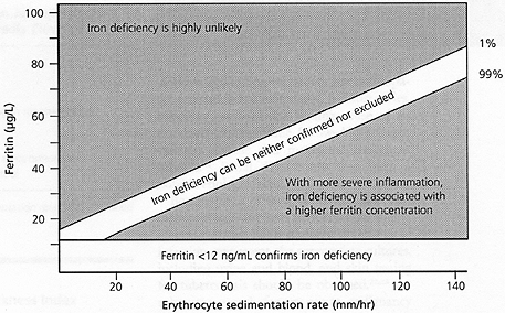
A nomogram to verify the presence or absence of iron deficiency coexistent with an underlying inflammatory condition by correlating serum ferritin level with degree of inflammation as evidenced by the erythrocyte sedimentation rate.
Reprinted with permission from Witte DL, Angstadt DS, Davis SH, Schrantz RD. Predicting bone marrow iron stores in anemic patients in a community hospital using ferritin and erythrocyte sedimentation rate. Am J Clin Pathol 1988;90:86.
Screening for Systemic Disease or Neoplasia
Unfortunately, the ESR is neither sensitive nor specific when used as a general screening test.1,3,19 For instance, the ESR may be elevated in the presence of infectious disease, other inflammatory or destructive processes, collagen vascular disease or malignancy,1–3 but it may not be increased in a number of infectious diseases (e.g., typhoid fever, malaria, mononucleosis), allergic processes, angina (as opposed to myocardial infarction) or peptic ulcer disease (as opposed to active inflammatory bowel disease).
Because an elevated ESR may occur in so many different clinical settings, this finding is meaningless as an isolated laboratory value. In addition, some patients who have malignant tumors, infections or other inflammatory disorders will have normal ESR values. Most unexplained ESR elevations are short-lived and not associated with any specific underlying process. In those instances where disease is present, it will usually be obvious after completion of history taking, physical examination and collection of routine laboratory data.3
Although an elevated ESR may occur with many types of cancer, it rarely indicates an occult tumor because most of these patients have widely metastatic disease.3,14,16 For this reason, when a mild to moderate elevation of the ESR (less than 100 mm per hour) is encountered in an asymptomatic patient, simply repeating the test at some future time should be considered in the absence of other clinical findings.3 No evidence suggests that an elevated ESR that is unsubstantiated by history, physical examination or other findings should trigger an extensive laboratory or radiographic work-up or invasive diagnostic procedures.1–3
Screening for Infection in Specific Clinical Settings
Recent studies have evaluated the ESR as a screening test for infection in specific clinical instances such as infection associated with orthopedic prostheses, pediatric bacterial infection and gynecologic inflammatory disease.6,7,20 Although frequently abnormal in patients who have an infected prosthesis, the ESR value is not as sensitive or specific an indicator of infection as joint aspiration.20 Elevation of the ESR has been proposed as a clue to the presence of an invasive bacterial infection in children after the first 48 hours of symptoms.6 In one investigation,7 the ESR more accurately indicated the severity of acute pelvic inflammatory disease than did the physical examination, thus helping to evaluate patients who required antimicrobial therapy. The appropriateness of the ESR as a screening test for infection, even in these well-defined clinical settings, requires further evaluation.
Usefulness as a Sickness Index in the Elderly
Some authors have proposed that the ESR be used as an inexpensive “sickness index” in the elderly.19,21 In a study of 142 residents of a long-term care hospital who had a nonspecific change in health status or developed new musculoskeletal complaints, the post-test probability of new disease rose from 7 percent in those with an ESR of less than 20 mm per hour to 66 percent in those with an ESR of more than 50 mm per hour. However, this investigation specifically excluded patients known to have an ESR-elevating disease and those in whom no disease was suspected.21
The authors concluded that combining clinical evaluation with an individual ESR value allowed the identification of groups of patients in whom the likelihood of disease was quite low or reasonably high, possibly limiting unnecessary investigations.
Extreme Elevation of the ESR
An extreme elevation of the ESR (defined as greater than 100 mm per hour) is associated with a low false-positive rate for a serious underlying disease.22,23 The conditions found in this situation have varied in individual populations, depending on patient age and inpatient versus outpatient status. In most series, infection has been the leading cause of an extremely elevated value, followed by collagen vascular disease and metastatic malignant tumors.22 Renal disease has also been a notable etiologic factor.3
Because most of these conditions are clinically apparent, any tests performed should be clinically driven. For instance, if symptoms of infection are present, the appropriate cultures, including urine and blood, and skin testing for tuberculosis should be obtained.22,23 An exhaustive search for an occult malignancy should not be undertaken because, if cancer is present, it is almost always metastatic.1,3
No obvious cause is apparent in fewer than 2 percent of patients with a markedly elevated ESR. In such patients, the history and physical examination coupled with readily available tests (Table 5) will usually establish the etiology. Because a notable number of patients with an ESR greater than 100 mm per hour have myeloma or some other type of dysproteinemia, urine and serum protein electrophoretic studies should be included in the testing.3
TABLE 5 Possible Testing in an Asymptomatic Patient with a Markedly Elevated ESR*
| PPD testing |
| Chest radiograph |
| Hematology profile |
| Creatinine and urea nitrogen measurements |
| Liver function studies |
| Urinalysis |
| Serum and urine protein electrophoresis |
| Occult blood testing of stool |
ESR = erythrocyte sedimentation rate; PPD = purified protein derivative.
*—> 100 mm per hr.
