Am Fam Physician. 2000;62(7):1474-1477
This feature is part of a year-long series of excerpts and special commentaries celebrating AFP's 50th year of publication. Excerpts from the two 1950 volumes of GP, AFP's predecessor, appear along with highlights of 50 years of family medicine.
This feature, titled “Office Pinch Grafting,” written by James L. Southworth, M.D., is reproduced from the April 1950 issue of GP. The commentary is provided by Clarissa Kripke, M.D., Assistant Clinical Professor, University of California, San Francisco, School of Medicine. Dr. Kripke is an assistant editor for AFP.
Transplantation of small pinch grafts can be quite a simple procedure, readily carried out in the office or dispensary. In favorable circumstances the healing of indolent wounds, ulcers, or burns may be aided and with a minimum of scarring. In many instances the period of healing may be materially shortened. The method of transferring pinch grafts herein described requires a minimum of time and equipment and is applicable to several conditions. …
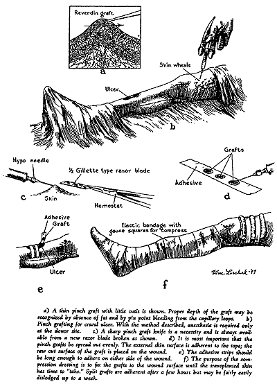
The depth of a pinch graft is shown in the accompanying plate (Figure a); this sort of graft is a Reverdin (1870) graft. Similar grafts may be cut deeper, extending into the cutis, but they are less sure of taking than the thinner graft illustrated.

Select a small area of skin which is of similar texture as the part to be grafted and in a convenient location. Shave and wash with soap, water, and alcohol. Plan to cut the grafts 0.5 cm in diameter and to place them about 0.5 cm apart on the wound. With 1 percent procaine and a fine sharp 25-gauge needle, raise skin wheals in the donor site—one for each graft (Figure c) and cut out a pinch of skin to the depth shown in Figure a. The proper depth can be recognized after a few trials by the fact that no subcutaneous fat can be seen if the graft is thin enough. If accidentally cut too deep, use the graft anyway—do not replace it in its bed. Lay the pinch grafts out in rows by sticking the external surface to a narrow flamed adhesive plaster strip (Figure d), taking care to straighten out the thin edges of the graft as shown in the figure. Transplant the pinch grafts in rows by sticking the adhesive plaster and its attached grafts across the wound (Figure e). Over the tape strips, place several dry gauze sponges or pads to form a resilient dressing. Apply a snug compression bandage as shown in Figure f.
If a graft is placed over a joint or on a finger or other mobile part, mobility must be eliminated by including an appropriate splint within the compression dressings. The donor site should be dressed with dry sterile gauze which is left in place two weeks.
These grafts may be redressed and examined as early as five days or as late as fourteen days: in any event the adhesive strips will have become loose so that there is little danger of pulling the new skin off. The overlying gauze, however, should be removed with care at the first dressing.—James L. Southworth, M.D.
Commentary
Although office pinch grafting is not used very frequently today, it is still a feasible technique that has a role in managing small burns, avulsed wounds and stasis ulcers. These small, full-thickness grafts are not as cosmetically appealing as split-thickness or synthetic-tissue grafts. They leave a cobblestone appearance at both the donor and the recipient site. However, they are effective and can be performed in any setting.
The technique for pinch grafts has not changed over the years. In his article on office pinch grafting for general practitioners, Dr. James Southworth describes the procedure. Pinch grafts are not hard to do. They are so easy in fact, that after reading a two-page article and looking at the illustration, I get the feeling that I could do one. The tone of the article demonstrates that Dr. Southworth thinks so too. He says, “The proper depth can be recognized after a few trials by the fact that no subcutaneous fat can be seen if the graft is thin enough. If accidentally cut too deep, use the graft anyway.” A needle, syringe, hemostat, half of a Gillette razor and a piece of tape are the only items of equipment needed.
Today, many graduates of family practice programs are not aware that tissue can be regenerated in this manner. The American Academy of Family Physicians still lists pinch grafts on the Privilege List for family practice departments in hospitals. However, the technique is no longer in the core curriculum list for residents. More often than not, skin grafts today are the domain of dermatologists and plastic surgeons. As technology advances, it is helpful to look backward every so often to rediscover the basics that we once knew.—clarissa kripke, m.d.