A 45-year-old woman presented with a one-year history of erythematous patches on her hands, chest, and upper back. She recently began having difficulty lifting her arms to brush her hair and climbing stairs.
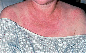
Skin examination revealed erythematous patches on the dorsal surface of the metacar-pophalangeal, proximal interphalangeal, and distal interphalangeal joints (Figure 1). Large areas of erythematous skin with telangiectases appeared on her upper chest, back, and shoulders (Figure 2).
FIGURE 1
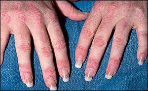
FIGURE 2
Question
Based on the patient's history and physical examination, which one of the following is the most likely diagnosis?
A. Porphyria cutanea tarda.
B. Scleroderma.
C. Systemic lupus erythematosus.
D. Polymyositis.
E. Dermatomyositis.
Discussion
The answer is E: dermatomyositis (DM), an inflammatory disease that causes characteristic skin changes as well as progressive, proximal muscle weakness. The average age of onset in adult patients is 40 years, but DM also can affect children and younger adults. It is more common in women and those with connective tissue diseases. It may be associated with a malignancy, particularly in patients who are 55 years or older.
The exact etiology of this disease is unknown, but an autoimmune cause is likely because of the presence of certain autoanti-bodies seen in patients with DM.1
The clinical presentation consists of skin findings and muscle weakness. The pathognomonic skin manifestation occurring in 70 percent of patients is the presence of erythematous patches on the skin overlying the dorsal surface of the metacarpophalangeal and interphalangeal joints, the knees and elbows, known as Gottron's papules. A less common but classic skin manifestation is the purplish-red heliotrope eruption on the eyelids with periorbital edema, which occurs in 30 to 60 percent of patients with DM. Large confluent areas of erythema over the upper chest, back, and shoulders—known as the “shawl” sign—may be seen, as in this patient. Periungual telangiectasias and generalized erythema on sun-exposed skin also occur with DM.
The signs of muscle involvement include progressive, symmetric proximal muscle weakness, and weakness of the pharyngeal and tongue muscles. Proximal muscle weakness is manifested by difficulty climbing stairs, rising from a seated position, or raising the arms overhead (e.g., brushing one's hair). It is not uncommon for the myositis to lead to serious complications if it affects the diaphragm, accessory respiratory muscles, or cardiac muscle. The pharyngeal muscle weakness may lead to dysphagia, aspiration, and respiratory tract infections. Progressive pulmonary fibrosis occurs in 10 percent of patients and, along with myositis of the respiratory muscles, may lead to hypoxemia. Cardiomyopathy may cause arrhythmias or heart failure. These complications are the major causes of mortality in patients with DM.1
The clinical presentation, enzyme elevations, electromyographic abnormalities, and the presence of myositis-associated autoantibodies are helpful tools in the diagnosis of DM. In DM, the amount of creatine kinase MM subtype fluctuates with disease activity.
Patients with DM may respond well to steroid treatment, and it is the first line of therapy. If muscle strength does not improve after several months of steroid treatment, other immunosuppressants may be considered, such as methotrexate, azathioprine, cyclophosphamide, or cyclosporine.
Porphyria cutanea tarda presents as bullae, vesicles, and milia on the dorsal hands and forearms but does not cause muscle weakness.
In scleroderma, the fingers become tapered over time. The skin is tight and smooth without erythematous patches.
Systemic lupus erythematosus may cause erythematous patches on the face or dorsal fingers, but the skin involvement is usually between the joints, rather than on top of the joints as in DM. Arthralgias, not muscle weakness, are the most common musculoskeletal complaint in lupus.
Polymyositis does lead to proximal muscle weakness, but it does not have the skin findings found in DM.
