The diagnosis of an abnormal fontanel requires an understanding of the wide variation of normal. At birth, an infant has six fontanels. The anterior fontanel is the largest and most important for clinical evaluation. The average size of the anterior fontanel is 2.1 cm, and the median time of closure is 13.8 months. The most common causes of a large anterior fontanel or delayed fontanel closure are achondroplasia, hypothyroidism, Down syndrome, increased intracranial pressure, and rickets. A bulging anterior fontanel can be a result of increased intracranial pressure or intracranial and extracranial tumors, and a sunken fontanel usually is a sign of dehydration. A physical examination helps the physician determine which imaging modality, such as plain films, ultrasonography, computed tomographic scan, or magnetic resonance imaging, to use for diagnosis.
Examination of a newborn's fontanels offers the physician a window into the infant's developing brain and general state of health. The word “fontanel” is derived from the Latin fonticulus and the Old French fontaine, meaning a little fountain or spring.1–3 The normal fontanel varies widely in shape and time of closure. The incidence of abnormal fontanel differs, depending on the abnormality and cause.
FIGURE 1.
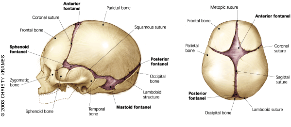
(Left) Lateral view of the newborn skull. (Right) Superior view of the newborn skull.
Redrawn with permission after Netter FH. Atlas of human anatomy. Summit, N.J.: Ciba-Geigy, 1994.
FIGURE 2.
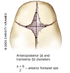
Measurement of the anterior fontanel.
Anatomy of the Fontanels
Fontanels are the fibrous, membrane-covered gaps created when more than two cranial bones are juxtaposed, as opposed to sutures, which are narrow seams of fibrous connective tissue that separate the flat bones of the skull.
A newborn has six fontanels (Figure 1): the anterior and posterior, two mastoid, and two sphenoid.4 The rhomboid-shaped anterior fontanel, located at the juncture of the two parietal and two frontal bones, is the most prominent. The superior sagittal dural venous sinus is partially situated beneath the anterior fontanel. The triangular posterior fontanel is located at the junction of the occipital and two parietal bones.1,5
Growth and Development of the Skull
The flat bones of the skull develop as part of the membranous neurocranium. Needle-like spicules radiate from a primary ossification center toward the periphery. These irregular bone islands are remodeled into flattened sheets of bone by osteoblast and osteoclast activity. During fetal and postnatal life, the membranous bones enlarge by resorption centrally and by apposition of new layers at the edges of the sutures.5
Growth of the cranium is triggered by brain growth, two thirds of which occurs by two years of age. Except for the metopic suture between the frontal bones, which closes at two years of age, the sutures remain open until brain growth ceases in the second decade of life.6 Once a suture is fused, growth perpendicular to that suture is restricted. Therefore, fontanel size is influenced by brain growth, dural attachments, suture development, and osteogenesis.7
Examination of the Fontanels
PHYSICAL EXAMINATION
The newborn's skull is molded during birth. The frontal bone flattens, the occipital bone is pulled outward, and the parietal bones override. These changes aid delivery through the birth canal and usually resolve after three to five days.8 The newborn's skull should be evaluated for shape, circumference, suture ridges, and size of anterior and posterior fontanels. Size is calculated by the average of the anteroposterior and transverse dimensions9 (Figure 2).
The fontanels should be examined while the infant is calm and held in both supine and upright positions. In select cases, such as newborns with multiple hemangiomas or heart failure, the anterior fontanel is auscultated to detect a bruit, which can indicate an arteriovenous malformation.10 Palpation of the fontanel in the upright position may reveal a normal, slight pulsation. If the fontanels are closed and intracranial pressure has increased, percussion produces a “cracked-pot” sound (dull, lacking resonance), known as Macewen's sign.
Any associated dysmorphic facial features should be noted. Asymmetry of the head is detected by looking at the infant's head from above. Head circumference is an important indicator of brain development and should be monitored over time, especially if a fontanel closes early.6,11
IMAGING
Plain radiographs of the skull are the least expensive way to evaluate the sutures and cranial bones, but they are limited by the lack of mineralization of the neonatal cranium. Bridging of bone over a suture, an indistinct suture, or sclerosis along the suture margins indicates fusion. Cortical thinning, widened sutures, and a beaten-metal appearance known as “thumbprinting” are associated with increased intracranial pressure.12
If the anterior fontanel is open, ultrasonography is useful to evaluate ventricular dilatation.13 A computed tomographic (CT) scan can detect a fused suture, dilated ventricles, enlarged subarachnoid space, brain size, or an intracranial or extracranial mass.14 Magnetic resonance imaging (MRI) can detect cortical and white-matter abnormalities, such as degenerative diseases, and document the extent of calvarial masses. Disadvantages of CT scans and MRI include cost, the need for sedation, and, in the case of CT, irradiation.13,15
Normal Fontanel
POSTERIOR FONTANEL
At birth, the average size of the posterior fontanel is 0.5 cm in white infants and 0.7 cm in black infants.16 The fontanel usually is completely closed by two months of age.10
ANTERIOR FONTANEL
The key feature of a normal anterior fontanel is variation. On the first day of an infant's life, the normal fontanel ranges from 0.6 cm to 3.6 cm, with a mean of 2.1 cm.17 Black infants have larger fontanels (1.4 cm to 4.7 cm).16 The fontanels of full-term and preterm infants are similar in size once preterm infants reach term. The fontanel can enlarge in the first few months of life,18 and the median age of closure is 13.8 months. By three months of age, the anterior fontanel is closed in 1 percent of infants; by 12 months, it is closed in 38 percent; and by 24 months, it is closed in 96 percent. Anterior fontanels tend to close earlier in boys than in girls; the initial size of the fontanel is not a predictor of when it will close.19
Abnormal Anterior Fontanel
LARGE FONTANEL AND DELAYED FONTANEL CLOSURE
A list of the medical conditions associated with a large fontanel or delayed fontanel closure can be found in Table 1.20,21 Achondroplasia, congenital hypothyroidism, Down syndrome, rickets, and increased intracranial pressure are among the most common conditions.
Achondroplasia is an autosomal-dominant disorder of the epiphyseal plate cartilage that results in dwarfism.22 At birth, the infant has an enlarged head, low nasal bridge, prominent forehead, and shortened extremities, in addition to a large fontanel.9
An elevated thyroid-stimulating hormone level on a newborn screening usually detects congenital hypothyroidism, but an abnormally large anterior fontanel in conjunction with an open posterior fontanel can be an early sign of the disorder. Myxedema and growth deficiency are later signs.
TABLE 1 Conditions Associated with an Enlarged Anterior Fontanel and Delayed Closure
| Conditions | Enlarged fontanel | Delayed closure | |
|---|---|---|---|
| Most common | |||
| Achondroplasia | ✓ | ✓ | |
| Congenital hypothyroidism | ✓ | ✓ | |
| Down syndrome | ✓ | ✓ | |
| Increased intracranial pressure | ✓ | ✓ | |
| Normal variation | ✓ | ✓ | |
| Familial macrocephaly | ✓ | ||
| Rickets | ✓ | ✓ | |
| Less common | |||
| Skeletal disorders | |||
| Acrocallosal syndrome (seizures, polydactyly, mental retardation) | ✓ | ||
| Apert's syndrome (craniosynostosis, proptosis, hypertension) | ✓ | ✓ | |
| Campomelic dysplasia (prenatal growth deficiency, large cranium, bowed legs) | ✓ | ||
| Hypophosphatasia (polyhydramnios, short, deformed limbs, soft skull) | ✓ | ✓ | |
| Kenny-Caffey syndrome (hypoparathyroidism, dwarfism, macrocephaly) | ✓ | ✓ | |
| Osteogenesis imperfecta (shortened limbs, wormian calvarial bones) | ✓ | ✓ | |
| Chromosomal abnormalities | |||
| Trisomy 13 (polydactyly, microcephaly, cleft lip and palate) | ✓ | ✓ | |
| Trisomy 18 (growth retardation, small cranium, open metopic suture) | ✓ | ✓ | |
| Congenital infections | |||
| Rubella (low birth weight, cataracts, “blueberry muffin” skin lesions) | ✓ | ✓ | |
| Syphilis (saddle nose deformity, joint swelling, maculopapular rash) | ✓ | ✓ | |
| Drugs and toxins | |||
| Aminopterin-induced malformation (craniosynostosis, absences of frontal bones, hypertelorism) | ✓ | ✓ | |
| Fetal hydantoin syndrome (microcephaly, broad nasal bridge, hypoplasia of nails) | ✓ | ✓ | |
| Dysmorphogenetic syndromes | |||
| Beckwith-Wiedemann syndrome (macrosomia, abdominal wall defect, macroglossia) | ✓ | ✓ | |
| Zellweger syndrome (high forehead, flat occiput, abnormal ears, hypotonia) | ✓ | ✓ | |
| Cutis laxa (pendulous skin folds, hoarse cry) | ✓ | ✓ | |
| VATER association (vertebral defects, anal atresia, tracheoesophageal fistula, renal dysplasia) | ✓ | ✓ | |
| Otopalatodigital syndrome (frontal bossing, broad terminal phalanges, syndactyly) | ✓ | ||
| Miscellaneous | |||
| Malnutrition (poor weight gain, asymmetric growth) | ✓ | ✓ | |
| Hydranencephaly (macrocephaly, thinned skull vault, primitive reflexes preserved) | ✓ | ||
| Intrauterine growth retardation (birth weight less than 2 standard deviations below mean) | ✓ | ||
A third fontanel between the anterior and posterior fontanels is associated with hypothyroidism and Down syndrome.23 Infants with Down syndrome often have a single palmar crease, flat occiput and facies, rounded ears, and slanted palpebral fissures.
Rickets resulting from vitamin D deficiency rarely occurs in the United States but is one of the five most common childhood diseases in developing nations. Risk factors include breastfeeding without vitamin D supplementation, dark skin, and low sunlight exposure. One of the signs of rickets is craniotabes, a softened outer table of the occipital bone that buckles under pressure, producing a reaction similar to a ping-pong ball indenting and popping back out. Craniotabes is not present at birth but develops over the first few months of life. Craniotabes can occur normally in premature infants and in children younger than six months.18,24,25 Disorders associated with increased intracranial pressure that results in an abnormally large fontanel or delayed fontanel closure are discussed later in this article.
SMALL FONTANEL OR EARLY FONTANEL CLOSURE
Fontanel closure that occurs as early as three months of age can be within normal limits, but careful monitoring of head circumference in such cases is essential to exclude a pathologic condition. The fontanel sometimes can be open but difficult to detect during a physical examination. Craniosynostosis and abnormal brain development are associated with a small fontanel or early fontanel closure.20
TABLE 2 Differential Diagnosis of Microcephaly
| Most common |
| Chromosomal defects |
| Congenital infections |
| Fetal alcohol syndrome |
| Hypoxic-ischemic encephalopathy |
| Normal genetic variation |
| Others |
| Autosomal dominant or recessive types |
| Dysmorphic syndromes |
| Malnutrition |
| Maternal phenylketonuria |
| Normal variation |
| Structural brain defects |
| Universal craniosynostosis |
Craniosynostosis is the premature closing of one or more cranial sutures, resulting in an abnormal head shape. The condition can be idiopathic or caused by hyperthyroidism, hypophosphatasia, rickets, or hyperparathyroidism.20 It is also associated with more than 50 syndromes, such as Apert's, Crouzon's and Pfeiffer's. The risk of primary isolated craniosynostosis is 0.4 per 1,000 live births, and the sagittal suture is most commonly involved.
Examination at birth of an infant with craniosynostosis might reveal a ridge over a suture or lack of movement along a suture when alternating sides are gently pressed. Overriding of sutures from the normal molding process should resolve within the first few days of life.9 Later physical findings in infants with primary craniosynostosis include stunted cranial growth, increased intracranial pressure, proptosis, strabismus, and hearing impairment.26
Plain radiographs of the skull are used for initial evaluation. If craniosynostosis is present, a three-dimensional CT scan is obtained to detect any underlying brain abnormalities and to assist planning for surgery.27
Abnormal brain development that results in microcephaly also can cause a small anterior fontanel or early fontanel closure. Prenatal trauma to the brain, such as maternal alcohol abuse, and postnatal trauma, such as hypoxia, are potential causes of microcephaly.20 Table 220,28 lists the differential diagnosis for microcephaly.
BULGING OR SUNKEN FONTANELS
Disorders associated with increased intracranial pressure can cause a bulging anterior fontanel. The most common disorders are meningitis, encephalitis, hydrocephalus, hypoxic-ischemic injury, trauma, and intracranial hemorrhage.20 Table 320 lists the differential diagnoses for a bulging fontanel. Palpation may reveal a tense fontanel that feels similar to bone.23
Meningitis and encephalitis also cause temperature instability, poor feeding, and irritability. If meningitis is suspected, a lumbar puncture should be performed to evaluate the cerebrospinal fluid for Gram stain, protein, glucose, cell count, and culture. A CT scan of a child with meningitis shows the subarachnoid space expanding into the anterior fontanel.21
Hydrocephalus can result from an imbalance between the production and the absorption of cerebral spinal fluid. This condition affects 3 per 1,000 live births. Most cases occur before two years of age, while the anterior fontanel is still open. Physical signs include an abnormal rate of head growth, frontal bossing of the forehead, widened sutures, and dilated scalp veins. Imaging with ultrasonography, CT, or MRI shows enlarged ventricles in the absence of brain atrophy. Because ultrasonic waves will not penetrate bone, the anterior fontanel must be open if ultrasonography is used for diagnosis.13,15
Hypoxic-ischemic injury results in cytotoxic edema and diffuse brain swelling. Associated findings include poor feeding, decreased muscle tone, respiratory difficulties, and alterations in consciousness. Intracranial hemorrhage can be intraventricular, parenchymal, subarachnoid, or subdural. Associated findings include decreased muscle tone, seizures, decreased hematocrit, vomiting, and alterations in consciousness.20
Tumors also should be considered in the differential diagnosis of a bulging fontanel. Dermoid tumors of the scalp are the most frequent lesions presenting over the anterior fontanel and also may be found over the posterior fontanel.29,30 They usually are slow-growing and nontender, and they are twice as common among girls. A CT scan is necessary to exclude intracranial involvement.30 Brain tumors, which can present with signs of increased intracranial pressure and focal neurologic findings, are best diagnosed with MRI.31
The primary cause of a sunken fontanel is dehydration. Other signs include reduced peripheral perfusion, poor skin turgor, and sunken eyes.32
TABLE 3 Differential Diagnosis of a Bulging Fontanel
| Hydrocephalus | |
| Space-occupying lesions | |
| Brain tumor | |
| Intracranial hemorrhage | |
| Brain abscess | |
| Infections | |
| Meningitis | |
| Encephalitis | |
| Roseola | |
| Shigella | |
| Mononucleosis | |
| Lyme disease | |
| Mastoiditis | |
| Cerebral malaria | |
| Cysticercosis | |
| Poliomyelitis | |
| Endocrine disorders | |
| Hyperthyroidism | |
| Hypoparathyroidism | |
| Pseudohypoparathyroidism | |
| Addison's disease | |
| Hypothyroidism | |
| Cardiovascular disorders | |
| Congestive heart failure | |
| Dural sinus thrombosis | |
| Hematologic disorders | |
| Polycythemia | |
| Anemia | |
| Leukemia | |
| Metabolic disorders | |
| Diabetic ketoacidosis | |
| Electrolyte disturbance | |
| Hepatic encephalopathy | |
| Uremia | |
| Galactosemia | |
| Hypophosphatasia | |
| Osteoporosis | |
| Maple syrup urine disease | |
| Miscellaneous | |
| Hypervitaminosis A | |
| Lead encephalopathy | |
| Aluminum toxicity | |
| Brain contusions | |
| Hypoxic-ischemic injury | |
| Coronal synostosis | |
| Trauma | |
| Dermoid cyst | |
Information from reference 20.
Final Comment
An abnormal fontanel in an infant can indicate a serious medical condition. Therefore, it is important to understand the wide variation of normal, how to examine the fontanels, and which diagnoses to consider when an abnormality is found. Consultation with a pediatric neurosurgeon should be considered if the diagnosis or presence of an abnormality is unclear.
