A 14-year-old male with type 1 diabetes presented to the emergency department complaining of sore throat, nausea, vomiting, and cough. He was treated with promethazine and amoxicillin, and discharged. He returned three days later with stomatitis, a finger-stick blood glucose level above 400 mg per dL (22.2 mmol per L), and serum ketones. He was admitted to the intensive care unit and given piperacillin-tazobactam, acyclovir, and fluconazole.
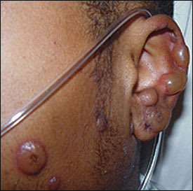
He had no known drug allergies or other recent drug exposures. Constitutional symptoms included weight loss of 8.1 kg (17.9 lb), fever, malaise, dyspnea, and a cough productive of yellow, blood-tinged sputum. Pertinent physical examination findings included a temperature of 40°C (104°F), a pulse of 138 beats per minute, and a respiratory rate of 23 breaths per minute. There was severe injection of the conjunctiva and sclera bilaterally and crusted lesions on the lips (Figure 1). Painful bullous lesions with petechia in the center were found on the ears (Figure 2), trunk, and extremities, including the palms and soles. A chest radiograph showed the presence of right lower lobe pneumonia.
Figure 1
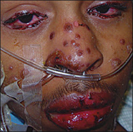
Figure 2
Question
Based on the patient’s history and physical examination, which one of the following is the correct diagnosis?
A. Viral exanthem.
B. Phototoxic eruption.
C. Toxic shock syndrome.
D. Stevens-Johnson syndrome.
E. Urticaria.
Discussion
The answer is D: Stevens-Johnson syndrome, caused by mycoplasma infection. After the antibiotic regimen was changed to azithromycin to treat the mycoplasmal pneumonia, the skin lesions crusted over, and the patient’s fever abated. After a 12-day hospital stay, the patient was discharged on oral azithromycin.
This patient presented with a mycoplasma infection, common in schoolchildren and adolescents, that progressed from a sore throat to pneumonia.1 The infection resulted in uncontrolled diabetes, erythema multiforme, and subsequent progression to Stevens-Johnson syndrome.
Erythema multiforme, Stevens-Johnson syndrome, and toxic epidermal necrolysis are thought to be a continuum of mucocutaneous reaction patterns of increasing severity.2 The underlying pathway is suspected to be an immunologic reaction in the skin, possibly triggered by immune complexes.
Erythema multiforme is a relatively common illness affecting predominantly younger patients. One half of cases occur in persons younger than 20 years, often after herpes or mycoplasma infections.3,4 The skin lesions start as macules that will blanch with pressure and later develop into classic target lesions that no longer blanch. Target lesions are diagnostic and have a central, dark-purple area or blister surrounded by a pale, edematous zone, which is in turn surrounded by a peripheral ring of erythema. These lesions tend to locate on the face and distal extremities, including the palms and soles. Lesions may be painful, especially those involving mucosal surfaces of the mouth, eyes, or genitals. Patients with erythema multiforme appear ill, with fever, prostration, and difficulty eating.
In patients who progress to Stevens-Johnson syndrome, there are more extensive skin and mucosal lesions. Mucous membrane involvement can be widespread, including the conjunctivae, nose, lips, oropharynx, tracheobronchial tree, genitalia, and anal tissue. Skin involvement also is more severe, with lesions moving in from the extremities to include the trunk. Eye involvement may spread beyond the conjunctivae, causing corneal ulcers or anterior uveitis. In Stevens-Johnson syndrome, erosions cover less than 10 percent of the body surface area. Tubular necrosis with acute renal failure can be noted. Stevens-Johnson syndrome is fatal in 5 percent of cases.
Toxic epidermal necrolysis is diagnosed when greater than 10 percent of the body surface is eroded. It does not always start with the typical target lesions but may begin with diffuse skin erythema that rapidly progresses to necrosis and skin detachment. Sheet-like loss of the epidermis is noted. There is a 20 to 30 percent mortality rate with toxic epidermal necrolysis, usually as a result of sepsis.
Viral exanthems are typically generalized cutaneous eruptions that can vary from maculopapular lesions to vesicles or pustules. Target-like lesions and mucosal involvement do not usually occur, and affected patients are not systemically ill.
Phototoxic eruptions are nonimmunologic reactions that resemble a sunburn. The rash appears two to six hours after sunlight exposure, in the presence of a photosensitizing substance such as tetracycline, tar, or the furocoumarins of some plant species.
Toxic shock syndrome is associated with infected wounds and tampon use. Skin findings include generalized erythroderma and hyperemia of the oral and genital mucosal surfaces. In more severe cases, petechiae and bullae may develop, but target lesions do not occur.
Urticaria is marked by transient large erythematous wheals, sometimes with central clearing. Lesions are pruritic and can be associated with angioedema. Patients with urticaria are not systemically ill.
Table 1 Selected Differential Diagnosis of Rash in an Adolescent
| Condition | Characteristics |
|---|---|
| Viral exanthem | Cutaneous eruptions varying from maculopapular to morbilliform to vesicular or pustular |
| Phototoxic eruption | Resembles sunburn appearing two to six hours after sunlight exposure in a photodistribution while using photosensitizing substances |
| Toxic shock syndrome | Diffuse erythroderma (sunburn-like rash), later desquamation of skin, particularly the palms and soles, and systemically ill |
| Stevens-Johnson syndrome | Skin and mucous membrane involvement; painful lesions start as macules then progress to target lesions that no longer blanch; lesions cover less than 10 percent body surface area |
| Urticaria | Large erythematous wheals, sometimes with central clearing; lesions are pruritic and can be associated with angioedema; individual lesions persist for less than 24 hours |
