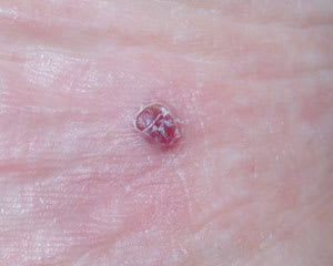
A healthy 56-year-old man presented with a painless nodular lesion on the plantar surface of his right foot (see accompanying figure). This nodule had grown slowly during the past year. The patient mentioned having a similar lesion one year ago that resolved spontaneously.
Question
Based on the patient’s history and physical examination, which one of the following is the correct diagnosis?
A. Pyogenic granuloma.
B. Nodular melanoma.
C. Eccrine poroma.
D. Classic Kaposi’s sarcoma.
E. Angiosarcoma.
Discussion
Answer is D: Kaposi’s sarcoma. Kaposi’s sarcoma is a low-grade, vascular neoplasm typically associated with human herpes virus 8 infection. The four clinical subtypes are classic, African, immunosuppression-associated, and acquired immunodeficiency syndrome (AIDS)–related Kaposi’s sarcoma. The morphology is clinically and histologically the same in all forms.
The classic form typically occurs in elderly men of Jewish, Italian, or Greek descent. It first appears on the toes or soles of the feet as violaceous macules and papules that slowly form plaques or rubbery-textured nodules. As the disease progresses, lymphedema may develop on affected extremities. The involvement of internal organs in this variant is rare. Spontaneous regression has been reported.1
African Kaposi’s sarcoma can be divided into cutaneous and lymphatic forms. The cutaneous form presents as nodules on the lower legs in middle-aged men. The lymphatic form occurs in children younger than 10 years and has a poor prognosis.
Immunosuppression-associated lesions resemble the classic form of Kaposi’s sarcoma, although they may be distributed in areas other than the feet or legs. These lesions may occur after solid-organ transplant and may progress to systemic involvement. Kaposi’s sarcoma is 300 times more common in immunosuppressed patients than in the population at large.2
AIDS-related Kaposi’s sarcoma is the most common tumor seen in patients with human immunodeficiency virus infection. It is 20,000 times more common in people with AIDS than in the general population.2 It presents most often on the lower extremities as painless red/violaceous papules or plaques that can be up to several centimeters in size. Systemic involvement is common.
The goal of treatment is to shrink the tumors, which may be achieved by cryotherapy, intralesional chemotherapy, and radiation therapy. Studies3,4 also have shown effective treatment using interferon alfa.
Pyogenic granuloma is a benign vascular proliferation presenting chiefly in children and young adults. It is a small (less than 1 cm), rapidly growing, dome-shaped mass of granulation tissue that can be sessile or pedunculated, often with a surrounding collarette of white scale. It occurs on exposed surfaces such as the hands, feet, and face, or at sites of previous trauma.
Nodular melanoma may present as red, brown, or black nodules. They comprise 15 to 20 percent of melanomas and often are misdiagnosed because they mimic other skin tumors. An amelanotic form of nodular melanoma is light beige in color.5 Biopsy usually establishes the correct diagnosis.
Eccrine poroma is a rare, soft, nontender, slow-growing tumor derived from an eccrine sweat gland that can become sessile or pedunculated. It occurs most often on the palmar and plantar surfaces, and can be verrucous or lobulated. It may ulcerate with occasional bleeding following minor trauma. The malignant counterpart of eccrine poroma is called porocarcinoma, and 50 percent of cases arise in preexisting eccrine poromas.6
Angiosarcomas are rare, soft tissue neoplasms. They occur most frequently in elderly men. Although angiosarcomas may occur in any location of the body, 50 percent of cases occur in the head and neck, and angiosarcomas of the lower extremities are the next most frequent type. Cutaneous angiosarcoma presents as an enlarging bruise, a blue-black nodule, or an unhealed ulceration. Bleeding and pain may be present.
Selected Differential Diagnosis of Nodular Lesions
| Condition | Characteristics |
|---|---|
| Angiokeratoma (localized) | Solitary red to blue-black hyperkeratotic vascular papule or nodule located on an extremity; multiple lesions may occur in association with certain genetically transmitted diseases. |
| Angiosarcoma | Enlarging bruise, blue-black nodule, or unhealed ulceration; bleeding and pain may be present. |
| Cherry angioma | Bright-red to violaceous dome-shaped vascular lesion occurring primarily on the trunk; may multiply and simulate petechiae; asymptomatic. |
| Classic Kaposi’s sarcoma | Violaceous macules and papules on the toes or soles of the feet; slowly the sarcoma progresses to rubbery-textured plaques. |
| Eccrine poroma | Soft, nontender, slow growing; occurs on the soles or palms; may be verrucous or lobulated. |
| Hemangioma | Soft bright-red to deep-purple nodule or plaque in children; does not blanch entirely on diascopy; may ulcerate and become secondarily infected with lesion regression. |
| Nodular melanoma | Red, brown, or black nodule; grows rapidly; may ulcerate or bleed. |
| Pyogenic granuloma | Dome-shaped mass of granulation tissue; may be sessile or pedunculated with surrounding collarette of white scale. |
