Surfers are prone to acute injuries as well as conditions resulting from chronic environmental exposure. Sprains, lacerations, strains, and fractures are the most common types of trauma. Injury from the rider’s own surfboard may be the prevailing mechanism. Minor wound infections can be treated on an outpatient basis with ciprofloxacin or trimethoprim-sulfamethoxazole. Jellyfish stings are common and may be treated with heat application. Other treatment regimens have had mixed results. Seabather’s eruption is a pruritic skin reaction caused by exposure to nematocyst-containing coelenterate larvae. Additional surfing hazards include stingrays, coral reefs, and, occasionally, sharks. Otologic sequelae of surfing include auditory exostoses, tympanic membrane rupture, and otitis externa. Sun exposure and skin cancer risk are inherent dangers of this sport.
Millions of surfers worldwide are prone to a unique constellation of acute and chronic conditions. Family physicians in coastal areas should be prepared to treat patients with surfing injuries such as envenomations, lacerations, sprains, and fractures, and to counsel surfers about the risks of sun exposure.
Strength of Recommendation
| Key clinical recommendation | Label | References |
|---|---|---|
| Heat is recommended to reduce the pain associated with jellyfish stings. | B | 8,9 |
A = consistent, good-quality patient-oriented evidence; B = inconsistent or limited-quality patient-oriented evidence; C = consensus, disease-oriented evidence, usual practice, or case series. See page 2237 for more information.
Trauma
Two studies1,2 assessed the frequency and types of surfing injuries. Sprains, dislocations, strains, lacerations, and fractures were found to be the most common injuries. One study2 found an overall rate of 3.5 injuries per 1,000 surfing days. More advanced surfers, who often engage larger waves in more extreme conditions, were found to have more severe injuries then less experienced surfers.
Safety measures can help reduce the frequency and severity of injuries. Most surfers are injured from contact with their own surf board’s side rails and fins (Figure 1).1,2 Several safety devices are available, but none has been proven to prevent surfing injuries. Rubber guards for the board’s nose and soft-edged or rubber-guarded fins may prevent lacerations without altering the dynamics of the surf board. Surfing helmets may prevent head injuries.3 Protective eyewear designed specifically for surfers may protect against ultraviolet rays and orbital trauma.
Figure 1.
Anatomy of a surfboard.
The use of a surfboard leash for injury prevention is controversial. Leashes seem to reduce the number of accidents caused by loose boards hitting other surfers,1 and they ensure that surfers will have access to a flotation device if they are injured seriously. However, a leash keeps the board near the surfer, and recoil from the leash increases the risk of injury. Ocular trauma most often occurs when the board’s nose strikes the surfer’s eye; one study implicates leash recoil as a cause.4,5 Leashes are sold in various lengths. Longer leashes may decrease recoil injury, but they can increase the risk of injury to others.
Lacerations can become infected with marine organisms. Common pathogens isolated from seawater and marine wounds include Streptococcus species, Escherichia coli, Pseudomonas aeruginosa, Mycobacterium marinum, Staphylococcus aureus, Vibrio cholerae, Vibrio vulnificus, and Vibrio parahaemolyticus.6 Wounds should be cultured for aerobic, anaerobic, and marine organisms. Physicians should alert laboratory personnel that marine organisms may be present, because specialized media or additional sodium chloride may be necessary to identify these pathogens properly.6
Minor wounds usually do not require antibiotic treatment. Serious wounds or wounds in immunocompromised patients warrant empiric antibiotic treatment.7 Initial outpatient therapy is directed at Vibrio species and includes ciprofloxacin (Cipro) or trimethoprim-sulfamethoxazole (TMP-SMX; Bactrim, Septra).6 Parenteral antibiotics that are appropriate for initial therapy include cefotaxime (Claforan), ceftazidime (Fortaz), chloramphenicol (Chloromycetin), gentamicin (Garamycin), and tobramycin (Tobrex). Lacerations should be allowed to heal secondarily or, if necessary, by delayed primary closure.7
Marine Hazards
ENVENOMATION
Although clinical presentation may vary, certain treatment principles apply to all types of marine envenomation. First, wounds can become infected and should be treated as discussed above. Second, the possibility of retained foreign bodies should be considered in most patients with envenomations. Depending on the mechanism of injury and level of clinical suspicion, investigation of a retained foreign body can be done through wound exploration or appropriate radiographs. Finally, tetanus immunization should be given, if necessary.6
COELENTERATES
Coelenterates are invertebrates and can be free-floating or sessile. Surfers more often encounter free-floating coelenterates such as the true jellyfish, Portuguese man-of-war, and box jellyfish. These animals have a main body and multiple dangling tentacles with venom-filled cells called nematocysts. Nematocysts inject toxins subcutaneously in response to chemical or mechanical stimuli. Local symptoms of nematocyst envenomation from jellyfish stings include burning pain, erythema, edema, urticaria, and bullae formation, all of which can progress to skin necrosis (Figure 2). Wet suits may thwart some envenomations by preventing the toxin from reaching the skin. However, stings through wet suits have been reported.
Figure 2.
Jellyfish stings
Initial treatment of envenomation involves preventing further toxin release by removing any remaining tentacles or other retained animal parts. There is no consensus for the best method of inactivating nematocysts and reducing pain. There is some evidence that immersion of the affected area in hot water and the application of heat packs are effective pain-relief methods.8,9 Other treatments recommended by published and anecdotal sources10–12 include application of cold packs and irrigation with ethanol, vinegar, urine, baking soda, or methylated spirits. There is insufficient evidence to support one treatment over others because of the variety of study designs and jellyfish species that were used. Vigorous rubbing and irrigation with fresh water are not recommended because these methods may induce additional nematocyst discharge.13
Further treatment should address local and systemic symptoms. Pain and dermatitis may be treated with analgesics and antihistamines. Patients with a systemic reaction may have multiple organ system involvement and present with a variety of symptoms (Table 1).14 Systemic symptoms can occur five minutes to several hours after exposure. Patients with systemic symptoms should be monitored for symptom rebound for at least six to eight hours.15
TABLE 1 Systemic Symptoms of Marine Envenomation
| Cardiovascular symptoms |
| Arrhythmias |
| Hypertension |
| Tachycardia |
| Gastrointestinal symptoms |
| Abdominal pain |
| Nausea |
| Vomiting |
| Immune symptoms |
| Anaphylaxis |
| Musculoskeletal symptoms |
| Arthralgias |
| Myalgias |
| Spasm |
| Neurologic symptoms |
| Ataxia |
| Coma |
| Headache |
| Paralysis |
| Seizures |
| Ocular symptoms |
| Conjunctivitis |
| Corneal abrasions and ulcers |
| Renal symptoms |
| Oliguria |
| Renal failure |
| Respiratory symptoms |
| Bronchospasm |
| Respiratory failure |
Information from reference 14.
The box jellyfish is particularly venomous (Figure 3). Hawaii is the only area in the United States where this type of jellyfish is found. Antivenin for the box jellyfish should be used if envenomation is confirmed or suspected.
SEABATHER’S ERUPTION
Seabather’s eruption occurs when a person is exposed to the larvae of certain coelenterates. This intensely pruritic rash is thought to be a hypersensitivity reaction to the larval toxin. It has been reported most often in Bermuda, the Caribbean region, and the eastern coast of the United States.16 It is thought that nematocysts discharge toxin as the larvae get trapped in the swimsuit. Further toxin release can occur when the bather rinses in fresh water.17
Patients with seabather’s eruption present with an urticarial maculopapular rash on areas of the body that were covered by the swimsuit. The rash may appear while the bather is in the water or up to one and one half days later. The rash may last for two to 28 days; most reactions resolve within one to two weeks.18 Systemic symptoms occur most often in children and may include fever, nausea, vomiting, and headache.17 Initial treatment involves the topical application of heat or vinegar. Further treatment is symptomatic and may include topical corticosteroids, oral antihistamines, and oral steroids. Twice-daily application of 1.5 g of thiabendazole (Mintezol) for two days has been reported as efficacious.19 The swimsuit should be cleaned thoroughly because larvae can persist and re-envenomate.
STINGRAYS
Stingrays are bottom-dwelling creatures that usually are encountered while surfers are entering or exiting the water. Their tail ensheathes a sharp spine that can penetrate wet suits and water shoes. Patients who have been stung may present with puncture wounds or lacerations, usually in the lower extremities, and pain disproportionate to the wound’s appearance.7 Initial treatment with hot-water immersion inactivates the heat-labile toxin. Retained animal parts should be excluded by wound exploration or radiographs. Shuffling the feet while walking through shallow water can prevent stings, because stingrays scatter when they are alerted to human presence.
CORALS
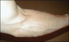
Coral reefs are common sources of lacerations and envenomations. Because of the variety of plant and animal species found on coral reefs, envenomation usually involves toxins from multiple sources. Patients with coral lacerations usually present with pain, pruritus, and erythema (Figure 4). Wounds should be irrigated and debrided, if necessary. Acetic acid has been recommended for treatment of stinging pain associated with coral envenomation.15 Wounds may heal slowly because of retained foreign material, nematocyst discharge, and microbial inoculation.20 Empiric antibiotic therapy as described above is appropriate if the wound appears to be infected.7
Figure 4.
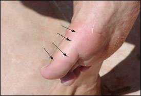
Coral lacerations on the foot of an adult male surfer. Sea urchin spines embedded in the lacerations also are visible (arrows).
SHARKS
Although shark attacks garner worldwide headlines, they are rare. In 2003, there were 55 unprovoked shark attacks worldwide, which resulted in four deaths.21 Of these, 39 attacks and one death occurred in the United States, mostly in Florida. About one half of all unprovoked shark attacks worldwide involved surfers or windsurfers.
Otologic Issues
AUDITORY EXOSTOSES
Auditory exostoses are bony outgrowths that arise from the temporal bone and protrude into the ear canal. Exostoses form in response to chronic exposure to cold water. They usually are asymptomatic, but patients can present with conductive hearing loss, frequent ear infections, and, occasionally, pain. Exostoses usually occur bilaterally, with multiple exostoses in a single ear canal.
The presence and severity of auditory exostoses directly correlates with the amount of time the patient spends in the water. Cold-water surfers are at higher risk for developing exostoses than warm-water surfers.22 Additional studies23,24show that persons who surf more frequently and for more years have a higher prevalence and severity of auditory exostoses than less experienced surfers. The consistent use of earplugs may help prevent exostoses from forming. Surgery is the only treatment for exostoses, and it usually is reserved for patients with severe, symptomatic cases.
OTITIS EXTERNA
Trauma, chronic exposure to moisture, and exostoses make otitis externa a common ailment among surfers. Otitis externa usually is caused by water stagnating in the external auditory canal. Trauma from foreign bodies and wave pressure also can contribute to infection.
Common infecting organisms include P. aeruginosa and S. aureus. Most patients can be treated empirically with topical antibacterial drops for five to seven days, although some patients may need treatment for up to two weeks.25 Systemic antibiotic therapy is recommended in patients with persistent cases or if otitis media also is present or suspected to have spread beyond the ear canal.25 Fungal species such as Aspergillus and Candida also may cause otitis externa, especially in patients with diabetes. Cultures or potassium hydroxide (KOH) preparations should be considered if antibacterial treatment fails. Preventive options include the use of earplugs while surfing and the routine use of isopropyl alcohol/acetic acid ear drops after surfing.
TYMPANIC MEMBRANE RUPTURE
Tympanic membrane rupture may occur when a surfer is struck by a strong wave or hits the water with sufficient force after a fall. Patients with tympanic membrane rupture may present with ear pain, conductive hearing loss, tinnitus, vertigo, and bloody otorrhea.26 Most ruptures heal spontaneously. Infection is common, so a short course of topical antibiotic therapy is indicated for surfing-related ruptures.27
Patients should be counseled to keep foreign material, including water, out of the ear and avoid surfing until the perforation heals. Molded earplugs may be used to keep water out of the ear during the healing process.22 Earplugs or helmets may reduce the likelihood of tympanic membrane rupture.
Sun Exposure
One small screening study28 found that surfers have an increased incidence of basal-cell skin cancer compared with a self-selected, age-matched control group. Sunscreen use reduces squamous-cell cancer; however, evidence for melanoma and basal-cell carcinoma is less clear. For example, melanoma risk may be related more closely to exposure intensity (i.e., sunburn) than the cumulative exposure. Persons using sunscreen may prolong sun exposure, thereby inadvertently increasing intensity and, thus, melanoma risk. Physicians can counsel patients about cancer risk and recommend using sunscreen with ultraviolet A and ultraviolet B protection, avoiding the sun between 10:00 a.m. and 4:00 p.m., and wearing protective clothing.29 It is unclear whether counseling patients about reducing their skin cancer risk actually leads to behavior change.
