
Am Fam Physician. 2005;72(6):1093-1094
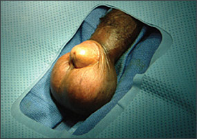
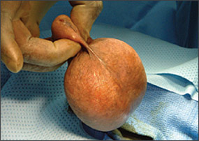
A 53-year-old man presented with a chief complaint of a “knot on my penis.” The patient related that the mass had been slowly increasing in size over the past several years. He was circumcised and denied any pain or difficulty with erection. He also denied any dysuria or urethral discharge. The patient was in a stable, monogamous relationship and had no known exposure to sexually transmitted diseases. His medical history was unremarkable, although he had not had a physical examination recently. He took no prescription, over-the-counter, or herbal medications. He had smoked one half of a pack of cigarettes per day for 27 years and denied use of alcohol. Review of systems was otherwise noncontributory. Physical examination revealed an obvious growth on the penis. The mass was smooth, round, uniform, doughy, and well circumscribed. It was approximately 8 cm in diameter and seemed to be attached to the distal shaft of the penis (Figures 1 and 2). The mass was removed surgically and sent for pathologic diagnosis.
Question
Discussion
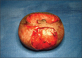
The answer is C: obstruction of smegma-producing glands. Surgical removal shows an encapsulated mass (Figure 3).
Smegma glands are sebaceous glands located within the inner surface of the foreskin.1,2 They appear as small white spots on either side of the frenulum at the point where it joins the glans. These white spots may be mistaken by physicians and uncircumcised patients as abnormal. They secrete a cheesy substance called smegma, a natural emollient of desquamated epithelial cells. Smegma serves to protect and lubricate the glans and inner lamella of the prepuce, facilitating erection, preputial eversion, and penetration during sexual intercourse. As with any sebaceous gland, smegma glands are subject to obstruction, which can lead to impressive enlargement given the anatomy of the soft tissues in this area. Their presence and potential for obstruction are not considered a medical indication for circumcision. Most, but not all, are removed by the circumcision procedure.
A penile carcinoma or malignant neoplasm would be firm in consistency, not doughy, and most likely would be fixed and nonpainful. Penile carcinoma is rare in the United States. It would have an insidious onset, as seen here. Penile lesions with an uncertain diagnosis often are biopsied or removed to confirm diagnosis and rule out carcinoma.
Paraphimosis is a painful swelling of the glans penis occurring when foreskin is left retracted. Paraphimosis would be circumferential (unlike the mass in Figure 3) and present more acutely. It is a vascular swelling rather than an encapsulated mass.
Condyloma would have a verrucous appearance and be more superficial. There may or may not be a history of exposure to sexually transmitted diseases. Although condyloma lesions can become large if untreated, their appearance usually is more characteristic with growth.
Penile edema often is preceded by infection or trauma and generally has a waxing and waning course. Simple edema would not be expected to be a worsening and chronic condition. Also, penile edema likely would be more diffuse and less defined.
| Condition | Characteristics |
|---|---|
| Penile carcinoma | Firm in consistency; likely to be fixed and nonpainful |
| Paraphimosis | Foreskin involvement; is circumferential |
| Obstruction of smegma-producing glands | Sebaceous glands located within the inner surface of the foreskin; doughy mass |
| Condyloma | Verruca-like appearance; superficial |
| Penile edema | Usually preceded by infection or trauma; waxing and waning course, not chronic |