Am Fam Physician. 2006;74(6):1011-1013
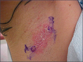
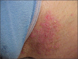
A 43-year-old woman presented with a recurrent rash in her axillae (Figure 1) and groin. She had a history of similar rashes occurring and resolving spontaneously over the previous 10 years. Her current eruption began about one month earlier in the right axilla and progressively involved the inframammary area and groin (Figure 2). She complained that the rash was extremely itchy, slightly malodorous, and worsened with heat. The patient recalled that her father and other members of her family had similar rashes. Examination revealed multiple macerated plaques with small, flaccid vesicles in the bilateral axillae, inguinal folds, and inframammary areas. The surrounding skin was erythematous and mildly tender to palpation. Cultures of the lesions grew group A streptococcus and Staphylococcus aureus.


Question
Discussion
The answer is C: Hailey-Hailey disease. Hailey-Hailey disease, or benign familial pemphigus, is a rare, chronic, autosomal dominant, inherited disorder initially described by Howard and Hugh Hailey in 1939.1 Patients with this disease often present with recurrent, tender, vesicular, crusted lesions in the intertriginous areas. The underlying cause of Hailey-Hailey disease stems from a mutation in the gene that encodes calcium adenosinetriphosphatase.2 Cellular (keratinocyte) adhesion in the midepidermis is then disrupted.
Hailey-Hailey disease usually appears in patients in their thirties and forties.1 Commonly affected areas include the neck, groin, axillae, and back. Patients often experience intense pruritus or a burning sensation that may be exacerbated by friction, heat, or sweating. Signs and symptoms tend to be worse in the summer than in the winter.1 Lesions can be described as an alternating crop of vesicles and erythematous plaques mixed with areas of dry crusts and erosions. Secondary bacterial, fungal, and viral infections are common. The lesions are malodorous and unsightly, causing significant distress and diminished social quality of life.
Diagnosis often is made clinically but may be confirmed by skin biopsy. In rare cases, asymptomatic longitudinal white bands on the fingernails may help in the diagnosis.1 Hailey-Hailey disease, unlike other forms of pemphigus, does not have an autoimmune basis, so biopsy immunofluorescence is negative. Although there is no definitive cure, topical corticosteroids and soothing compresses will provide some symptomatic relief. Topical or oral antibiotic and antifungal medications also may be used to treat secondarily infected lesions. Systemic corticosteroids have been used to control occasional f lares.2 Cyclosporine (Sandimmune), methotrexate, oral retinoids (e.g., acitretin [Soriatane]), dapsone, and tacrolimus (Prograf) may be effective in refractory cases, and dermatology consultation should be considered.2,3
Pemphigus vulgaris is a rare, autoimmune, blistering disease of the skin and the mucous membranes. It tends to affect persons in their fifties and sixties.4 Typically, the disease begins as oral mucosal sloughing and can spread quickly until all areas of the body are affected. Slight pressure or rubbing can cause skin separation.4 Immunofluorescent staining of biopsy specimens also can confirm the presence of intracellular autoantibodies.
Eczema herpeticum is caused by cutaneous herpes simplex virus infection in patients with atopic dermatitis. It is also called Kaposi’s varicelliform eruption.5 Patients typically present with clusters of umbilicated vesicles superimposed on active atopic damage. These lesions spread quickly, and the vesicles become small “punched out” erosions that can be infected secondarily. Patients may experience fever, malaise, and lymphadenopathy. Diagnosis is clinical, although Tzanck test, direct fluorescent antibody test, or viral culture can be used to confirm the diagnosis.
Intertrigo is a superficial inflammatory dermatitis affecting the top layers of the skin.4 It commonly arises in areas where two skin surfaces constantly rub or press against one another (e.g., skinfolds). Intertrigo is more prevalent in obese persons and can be exacerbated by hot and humid weather. The lesions can be a combination of erosions, fissures, and exudation, and the affected area may be infected secondarily, particularly by Candida.
Hidradenitis suppurativa is a chronic inflammatory condition typically affecting areas with apocrine glands, particularly the axillae, perineum, buttocks, and periareolar and perianal regions. The disease is thought to stem from a combination of genetic factors, excessive perspiration, hormones, and infection of the hair follicles.6 Patients commonly present with recurrent boils that can develop subsequently into sterile abscesses, sinus tracts, and fistulas.
| Condition | Characteristics |
|---|---|
| Contact dermatitis caused by deodorants | Inflammatory response of the skin to deodorant components such as cinnamic aldehyde (cinnamal), hydroxycitronellal, farnesol, and aluminum chloride |
| Eczema herpeticum | Cutaneous herpes simplex virus infection in patients with atopic dermatitis; clusters of umbilicated vesicles on atopic skin |
| Hailey-Hailey disease | Alternating crop of vesicles and erythematous plaques mixed with areas of dry crusts and erosions; favors intertriginous areas; genetic mutation |
| Hidradenitis suppurativa | A chronic inflammatory condition that typically affects areas of the skin bearing apocrine glands |
| Intertrigo | Superficial inflammatory dermatitis; commonly arises on opposing skinfolds; often secondarily infected |
| Pemphigus vulgaris | Rare autoimmune blistering disease; affects skin and mucous membranes |
| Tinea cruris | A pruritic superficial fungal infection of the groin and adjacent skin |