Granuloma annulare is a benign, asymptomatic, self-limited papular eruption found in patients of all ages. The primary skin lesion usually is grouped papules in an enlarging annular shape, with color ranging from flesh-colored to erythematous. The two most common types of granuloma annulare are localized, which typically is found on the lateral or dorsal surfaces of the hands and feet; and disseminated, which is widespread. Localized disease generally is self-limited and resolves within one to two years, whereas disseminated disease lasts longer. Because localized granuloma annulare is self-limited, no treatment other than reassurance may be necessary. There are no well-designed randomized controlled trials of the treatment of granuloma annulare. Treatment recommendations are based on the patho-physiology of the disease, expert opinion, and case reports only. Liquid nitrogen, injected steroids, or topical steroids under occlusion have been recommended for treatment of localized disease. Disseminated granuloma annulare may be treated with one of several systemic therapies such as dapsone, retinoids, niacinamide, antimalarials, psoralen plus ultraviolet A therapy, fumaric acid esters, tacrolimus, and pimecrolimus. Consultation with a dermatologist is recommended because of the possible toxicities of these agents.
SORT: KEY RECOMMENDATIONS FOR PRACTICE
| Clinical recommendation | Evidence rating | References |
|---|---|---|
| Localized granuloma annulare is self-limited and asymptomatic and usually does not require treatment. | C | 1 |
| Options for treatment of localized granuloma annulare include liquid nitrogen, injected steroids, and topical steroids. | C | 18 |
| Treatment for disseminated granuloma annulare should be undertaken in consultation with a dermatologist; options include dapsone, retinoids, antimalarial drugs, tacrolimus (Protopic), and pimecrolimus (Elidel). | C | 19–27,35,36 |
A = consistent, good-quality patient-oriented evidence; B = inconsistent or limited-quality patient-oriented evidence; C = consensus, disease-oriented evidence, usual practice, expert opinion, or case series. For information about the SORT evidence rating system, see page 1666 orhttps://www.aafp.org/afpsort.xml.
Granuloma annulare is a benign skin condition that typically consists of grouped papules in an enlarging annular shape. Their appearance ranges from flesh colored to erythematous. The etiology is unknown, but the disease usually is self-limited. Despite the dramatic appearance of this cutaneous eruption, it generally is asymptomatic; however, there may be some mild pruritus. The eruption can occur anywhere on the body, but it occurs least often on the face and most often on the lateral or dorsal surfaces of the hands and feet (Figure 1).
Figure 1.
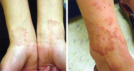
Localized granuloma annulare in a 25-year-old woman who has had the eruption on her (A) wrists and (B) ankles since age 17. The eruption spontaneously resolved but then recurred.
Epidemiology
Granuloma annulare affects patients of all ages. Most cases of localized granuloma annulare are diagnosed in patients before 30 years of age. Incidence is highest in women, with a ratio of 2.3 to 1.0 over men.1 Approximately 15 percent of all patients with granuloma annulare will have more than 10 lesions (i.e., disseminated granuloma annulare). These patients are usually children younger than 10 years or adults older than 40 years. Although uncommon, cases of granuloma annulare occurring in siblings, twins, and successive generations have been reported.2 Seasonal peaks of granuloma annulare in the spring and fall also have been described.3
The duration of the skin eruption varies. In more than one half of patients, it resolves spontaneously within two months to two years. However, cases of disseminated granuloma annulare may last three to four years or as long as 10 years. The eruption may recur as well, with 40 percent of children having recurrent lesions.4
Etiology
The cause of granuloma annulare is unknown, but it has been reported to follow trauma, malignancy, viral infections (including human immunodeficiency virus [HIV], Epstein-Barr virus, and herpes zoster), insect bites, and tuberculosis skin tests.5 A delayed-type hypersensitivity reaction and cell-mediated immune response are hypothesized. In one retrospective study, 12 percent of patients with granuloma annulare had diabetes mellitus.6 This study did not have a comparison group, so it is not clear whether the prevalence of diabetes mellitus was higher or lower than in the general population. Patients with diabetes mellitus had a higher incidence of chronic relapsing granuloma annulare than patients without diabetes. A case-control study that included patients with and without diabetes failed to reveal any statistically significant correlation between granuloma annulare and type 2 diabetes.7 Some isolated cases of granuloma annulare found in association with malignant neoplasm have been reported. In these cases, the malignant neoplasms were primarily lymphoma,8 but some were prostate cancer.9 Granuloma annulare has occurred in all stages of HIV infection as well.10
Clinical Presentation
The four main clinical variants of granuloma annulare are: localized, disseminated, subcutaneous, and perforating.
LOCALIZED
The localized form of granuloma annulare composes 75 percent of cases.11 Localized granuloma annulare starts as a ring of small, firm, flesh-colored or red papules. As the condition progresses, there is some central involution, and the ring of papules slowly increases from 0.5 to 5.0 cm in diameter. The lesions may be isolated or coalesce into plaques. They are found on the lateral or dorsal surfaces of the hands and feet (Figure 2). More than 50 percent of these patients will have spontaneous resolution within two years.5
Figure 2.
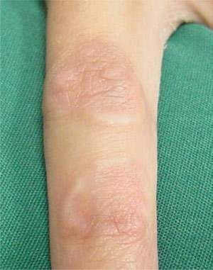
Localized granuloma annulare in a 27-year-old woman with an asymptomatic eruption for one year on her left index finger.
DISSEMINATED OR GENERALIZED
Disseminated or generalized granuloma annulare is similar to the localized variant but is more widespread, having 10 or more lesions (Figure 3). The papules may fuse to form annular lesions on the extremities, trunk, and neck. In contrast to the localized form, these lesions may persist for three to four years or longer.
Figure 3.
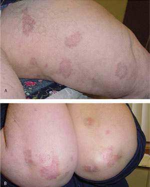
Disseminated granuloma annulare in a 62-year-old woman on her (A) inner thigh and (B) elbows. The eruption was asymptomatic.
SUBCUTANEOUS
Subcutaneous granuloma annulare is diagnosed primarily in children two to five years of age. The lesions are asymptomatic, rapidly growing subcutaneous nodules on the extremities, hands, scalp, buttocks, and pretibial and periorbital areas. The lesions may be solitary or in clusters. Diagnosis is made with an excisional biopsy. These lesions may resolve spontaneously or may recur after excision. There have been no reports of progression to systemic illness.12
PERFORATING
Perforating granuloma annulare is rare and occurs most often in children and young adults. It is also more common in women. Perforating granuloma annulare can have localized and generalized forms. The localized form is found on the upper limbs and pelvis, and the generalized form, which is more common, is present on the abdominal area, trunk, and upper and lower limbs. The lesions are 1- to 4-mm papules with a central crust or scale with or without an umbilicated center. Biopsy shows palisading granuloma with transepithelial elimination of degenerating collagen fibers.13 This transepithelial elimination leads to the perforating designation. Twenty-five percent of patients report pruritus, and 25 percent report pain, mainly in lesions located on the palms.14
Differential Diagnosis
Granuloma annulare can be mistaken for other common annular skin conditions such as tinea corporis, pityriasis rosea, nummular eczema, psoriasis, or erythema migrans of Lyme disease. The lack of any surface changes to the skin is the key feature that distinguishes granuloma annulare from these other skin conditions. Specifically, there is no scale or associated vesicles or pustules with granuloma annulare; the skin surface is smooth. Less common annular skin conditions (e.g., subacute cutaneous lupus erythematosus, erythema annulare centrifugum) have associated scaling and can be ruled out. Urticaria also can present as annular plaques, but it is distinguished easily from granuloma annulare by its evanescent nature.
A less common annular skin condition, sarcoidosis, may present with reddish-brown to purplish infiltrated papules and plaques that commonly are found on the face. Hansen's disease (leprosy) also has erythematous annular plaques with associated scaling, alopecia, and anesthesia.15
Often, a diagnosis can be made without a punch biopsy, but in clinically confusing situations it may be helpful, especially with the subcutaneous variant of granuloma annulare. The presence of epithelioid histiocytes palisading around an anuclear dermis with mucin deposition is characteristic of granuloma annulare. This granulomatous appearance of the biopsy and the annular clinical appearance combine to form the descriptive term “granuloma annulare.” If a biopsy is performed, the results typically will show focal degeneration of collagen with reactive inflammation and fibrosis.16 The epidermis is normal.
Treatment
Medical literature contains limited reliable information on the treatment of granuloma annulare. The only double-blind, placebo-controlled crossover study concerning the treatment of disseminated granuloma annulare involved the use of oral potassium iodide. In this series of eight patients, there was no advantage of high-dose potassium iodide over placebo.17
Most medical literature on treatment of granuloma annulare is limited to individual case reports and small series of patients treated without a control group. Such studies cannot establish treatment effectiveness, particularly with a self-limited disease.
Because localized granuloma annulare is self-limited and asymptomatic, treatment usually is not necessary. Nevertheless, many patients remain troubled by the appearance and persist in seeking treatment. For patients insisting on treatment, options include intralesional corticosteroid injection with 2.5 to 5.0 mg per mL triamcinolone (Aristocort) into the elevated border, topical corticosteroids under occlusion, cryotherapy, and electrodesiccation. Patients should be warned that all of these treatments could cause scarring and atrophy. One uncontrolled study of 31 patients with localized granuloma annulare showed 81 percent resolution after one treatment with liquid nitrogen or nitrous oxide.18
Systemic therapy is required for disseminated granuloma annulare, and many different treatments have been proposed (Table 117,19–37). The possible benefit of treatment, which is unclear given the lack of clinical trials, must be balanced against the significant toxicities of most of these treatments. Therefore, the family physician must proceed with caution and should consider consultation with a dermatologist.
TABLE 1 Summary of Studies on Treatment of Disseminated Granuloma Annulare
| Treatment type | Number of patients | Dosage and duration | Outcome | Side effects | ||
|---|---|---|---|---|---|---|
| Dapsone | ||||||
| Steiner, 198519 | 10 | 100 mg daily for 2 to 18 weeks | Four had complete resolution, three had partial response | Headache or weakness | ||
| Czarnecki, 198620 | 6 | 100 mg daily for 4 to 12 weeks | All resolved | Fatigue | ||
| Saied, 198021 | 2 | 100 to 200 mg daily for 4 to 44 weeks | One had complete resolution, one was resolving | None | ||
| Isotretinoin (Accutane) | ||||||
| Schleicher, 198522 | 1 | 40 mg once to twice daily for 12 weeks | 90 percent resolution | Dry lips, elevated triglyceride levels | ||
| Tang, 199623 | 1 | 30 to 50 mg daily for 16 weeks | Complete response | None | ||
| Buendia-Eisman 200324 | 1 | 50 mg daily for eight weeks | 90 percent resolution | None | ||
| Schleicher, 199225 | 7 | 40 mg daily for 10 weeks | 100 percent response; three recurred after initial clearing, and drug discontinued | Elevated liver function test results | ||
| Etretinate (not available in the U.S.) | ||||||
| Botella-Estrada, 199226 | 1 | 50 mg daily for 28 weeks | 90 percent resolution | Hair loss | ||
| Hydroxychloroquine (Plaquenil)/chloroquine (Aralen) | ||||||
| Carlin, 198727 | 1 | Hydroxychloroquine 200 mg twice daily for 12 weeks | Near complete clearing | None | ||
| Simon, 199428 | 1 | Two hydroxychloroquine, 6 mg per kg daily for six weeks | Complete clearing | None | ||
| Four chloroquine, 3 mg per kg daily for six weeks | ||||||
| Cyclosporine (Sandimmune) | ||||||
| Fiallo, 199829 | 2 | 3 mg per kg daily for 12 weeks | Complete clearing | None | ||
| Niacinamide | ||||||
| Ma, 198330 | 1 | 1,500 mg daily for 24 weeks | Complete clearing | None | ||
| Psoralen plus ultraviolet A (PUVA) | ||||||
| Setterfield, 199931 | 1 | 53 PUVA treatments | Complete clearing | None | ||
| Kerker, 199032 | 5 | 21 to 95 treatments | Complete clearing | None | ||
| Vitamin E/zileuton (Zyflo) | ||||||
| Smith, 200233 | 3 | Vitamin E, 400 IU daily | Complete clearing | None | ||
| Zileuton, 600 mg daily for eight to 12 weeks | ||||||
| Fumaric acid esters | ||||||
| Eberlein-Konig, 200534 | 8 | Standard therapy used for psoriasis | Four had complete remission, three had partial remission. | Diarrhea, dizziness, nausea | ||
| Topical tacrolimus 0.1% ointment (Protopic) | ||||||
| Harth, 200435 | 4 | Apply twice daily for eight weeks | Two patients had healing of inflammation. | Burning, itching | ||
| Topical pimecrolimus 1% cream (Elidel) | ||||||
| Rigopoulous, 200536 | 1 | Apply twice daily for 12 weeks | Partial clearing | None | ||
| Potassium iodide | ||||||
| Smith, 199417 | 8 | 3 to 10 drops three times dailyfor 24 weeks | No benefit over placebo | Rhinorrhea, metallic taste, acneform eruption | ||
| Infliximab (Remicade), a tumor necrosis factorαinhibitor | ||||||
| Hertl, 200537 | 1 | 5 mg per kg intravenously at 0, 2, and 6 weeks and monthly for four months | Near complete clearing | None | ||
IU = international units
Dapsone is an antibiotic commonly used for dermatitis herpetiformis or Hansen's disease. It has been reported to be effective in managing disseminated granuloma annulare.19–21 Isotretinoin (Accutane) is better known for treating severe acne, but it has been shown to be effective in treating granuloma annulare in numerous case reports.22–25 Serious adverse effects such as elevated triglyceride levels, elevated liver enzyme levels, and teratogenicity can occur. Two isotretinoin treatment failures also have been published.38 Etretinate, another retinoid (not available in the U.S.), also has been reported to be effective.26
Antimalarial agents, including hydroxychloroquine (Plaquenil) and chloroquine (Aralen), have been used in the treatment of granuloma annulare. They have been presumed effective because of their immunosuppressive and anti-inflammatory properties.27,28 Serious side effects are possible, including retinopathy, aplastic anemia, and liver toxicity. Effective use of cyclosporine (Sandimmune) has been reported in individual patients. 29 Close monitoring of serum creatinine levels and blood pressure is needed with this drug.
Niacinamide has been used and is reasonably safe, even at high doses. Nevertheless, liver toxicity is an important adverse effect, and hepatic transaminase levels should be monitored during treatment.30 Oral psoralen (e.g., anthralin [Anthra-Derm]) and psoralen plus ultraviolet A (PUVA) therapy has been reported to be effective in two uncontrolled studies with a total of six patients. However, long-term PUVA therapy carries a risk of increased incidence of nonmelanoma skin cancer.31,32 Vitamin E combined with a 5-lipoxygenase inhibitor (e.g., zileuton [Zyflo]) has been tried and was successful, but only in a series of three patients.33
Fumaric acid esters, which also are used to manage psoriasis, were found to have some benefit in a recent study treating eight patients. One half of the study participants discontinued therapy because of gastrointestinal side effects.34
In recent case reports, topical tacrolimus and pimecrolimus had positive outcomes. The incidence of side effects is very low.35,36
Infliximab (Remicade), a tumor necrosis factor B inhibitor, demonstrated a positive outcome in a patient with recalcitrant disseminated granuloma annulare.37 Granuloma annulare is difficult to treat clinically; reassurance that the condition will self-resolve may be the best option. Clearly, well-designed clinical trials are needed to better direct treatment.
