Impetigo is a highly contagious, superficial skin infection that most commonly affects children two to five years of age. The two types of impetigo are nonbullous impetigo (i.e., impetigo contagiosa) and bullous impetigo. The diagnosis usually is made clinically, but rarely a culture may be useful. Although impetigo usually heals spontaneously within two weeks without scarring, treatment helps relieve the discomfort, improve cosmetic appearance, and prevent the spread of an organism that may cause other illnesses (e.g., glomerulonephritis). There is no standard treatment for impetigo, and many options are available. The topical antibiotics mupirocin and fusidic acid are effective and may be superior to oral antibiotics. Oral antibiotics should be considered for patients with extensive disease. Oral penicillin V is seldom effective; otherwise there is no clear preference among antistaphylococcal penicillins, amoxicillin/clavulanate, cephalosporins, and macrolides, although resistance rates to erythromycin are rising. Topical disinfectants are not useful in the treatment of impetigo.
Impetigo is a highly contagious infection of the superficial epidermis that most often affects children two to five years of age, although it can occur in any age group. Among children, impetigo is the most common bacterial skin infection and the third most common skin disease overall, behind dermatitis and viral warts.1,2 Impetigo is more common in children receiving dialysis.1 The infection usually heals without scarring, even without treatment. Staphylococcus aureus is the most important causative organism. Streptococcus pyogenes (i.e., group A beta-hemolytic streptococcus) causes fewer cases, either alone or in combination with S. aureus.3
SORT: KEY RECOMMENDATIONS FOR PRACTICE
| Clinical recommendation | Evidence rating | References |
|---|---|---|
| Topical antibiotics such as mupirocin (Bactroban) and fusidic acid (not available in the United States) are the preferred first-line therapy for impetigo involving limited body surface area. | A | 4,8 |
| Oral antibiotics (e.g., antistaphylococcal penicillins, amoxicillin/clavulanate [Augmentin], cephalosporins, macrolides) are effective for the treatment of impetigo; erythromycin is less effective. | A | 4,8 |
| Oral antibiotics should be considered for patients with impetigo who have more extensive disease and for disease associated with systemic symptoms. | C | 4,8 |
| Oral penicillin V, amoxicillin, topical bacitracin, and neomycin are not recommended for the treatment of impetigo. | B | 4,8 |
| Topical disinfectants such as hydrogen peroxide should not be used in the treatment of impetigo. | B | 4,8 |
A = consistent, good-quality patient-oriented evidence; B = inconsistent or limited-quality patient-oriented evidence; C = consensus, disease-oriented evidence, usual practice, expert opinion, or case series. For information about the SORT evidence rating system, see page 789 orhttps://www.aafp.org/afpsort.xml.
There are two types of impetigo: nonbullous (i.e., impetigo contagiosa) and bullous. Nonbullous impetigo represents a host response to the infection, whereas a staphylococcal toxin causes bullous impetigo and no host response is required to manifest clinical illness.3 The diagnosis usually is made clinically and can be confirmed by Gram stain and culture, although this is not usually necessary. Culture may be useful to identify patients with nephritogenic strains of S. pyogenes during outbreaks of poststreptococcal glomerulonephritis or those in whom methicillin-resistant S. aureus is suspected.3
Epidemiology
Impetigo usually is transmitted through direct contact. In a study in the United Kingdom, the annual incidence of impetigo was 2.8 percent in children up to four years of age and 1.6 percent among children five to 15 years of age.4 Nonbullous impetigo accounts for approximately 70 percent of cases. Patients can further spread the infection to themselves or others after excoriating an infected area. Infections often spread rapidly through schools and day care centers. Although children are infected most often through contact with other infected children, fomites also are important in the spread of impetigo. The incidence is greatest in the summer months, and the infection often occurs in areas with poor hygiene and in crowded living conditions.1,3
Diagnosis
NONBULLOUS IMPETIGO
Nonbullous impetigo begins as a single red macule or papule that quickly becomes a vesicle. The vesicle ruptures easily to form an erosion, and the contents dry to form characteristic honey-colored crusts that may be pruritic (Figures 1 and 2). Impetigo often is spread to surrounding areas by autoinoculation. This infection tends to affect areas subject to environmental trauma, such as the extremities or the face. Spontaneous resolution without scarring typically occurs in several weeks if the infection is left untreated.5
Figure 1.

Nonbullous impetigo on the face.
Figure 2.
Nonbullous impetigo in the groin.
A subtype of nonbullous impetigo is common (or impetiginous) impetigo, also called secondary impetigo. This can complicate systemic diseases, including diabetes mellitus and acquired immunodeficiency syndrome. Insect bites, varicella, herpes simplex virus, and other conditions that involve breaks in the skin predispose patients to the formation of common impetigo. The presentation is similar to that of primary nonbullous impetigo. 1 Table 1 provides a selected differential diagnosis of nonbullous impetigo.1
TABLE 1 Selected Differential Diagnosis of Nonbullous Impetigo
| Diagnosis | Distinguishing features |
|---|---|
| Atopic dermatitis | Chronic or relapsing pruritic lesions and abnormally dry skin; flexural lichenification is common in adults; facial and extensor involvement is common in children |
| Candidiasis | Erythematous papules or red, moist plaques; usually confined to mucous membranes or intertriginous areas |
| Contact dermatitis | Pruritic areas with weeping on sensitized skin that comes in contact with haptens (e.g., poison ivy) |
| Dermatophytosis | Lesions may be scaly and red with slightly raised “active border” or classic ringworm; or may be vesicular, especially on feet |
| Discoid lupus erythematosus | Well-defined plaques with adherent scale that penetrates into hair follicles; peeled scales have “carpet tack” appearance |
| Ecthyma | Crusted lesions that cover an ulceration rather than an erosion; may persist for weeks and may heal with scarring as the infection extends to the dermis |
| Herpes simplex virus | Vesicles on an erythematous base that rupture to become erosions covered by crusts, usually on the lips and skin |
| Insect bites | Papules usually seen at site of bite, which may be painful; may have associated urticaria |
| Pemphigus foliaceus | Serum and crusts with occasional vesicles, usually starting on the face in a butterfly distribution or on the scalp, chest, and upper back as areas of erythema, scaling, crusting, or occasional bullae |
| Scabies | Lesions consist of burrows and small, discrete vesicles, often in finger webs; nocturnal pruritus is characteristic |
| Sweet's syndrome | Abrupt onset of tender or painful plaques or nodules with occasional vesicles or pustules |
| Varicella | Thin-walled vesicles on an erythematous base that start on trunk and spread to face and extremities; vesicles break and crusts form; lesions of different stages are present at the same time in a given body area as new crops develop |
Information from reference 1.
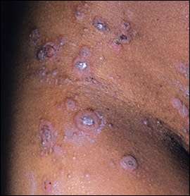
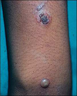
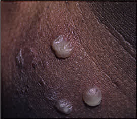
BULLOUS IMPETIGO
Bullous impetigo most commonly affects neonates but also can occur in older children and adults. It is caused by toxin-producing S. aureus and is a localized form of staphylococcal scalded skin syndrome.5,6 Superficial vesicles progress to rapidly enlarging, flaccid bullae with sharp margins and no surrounding erythema (Figures 3 and 4). When the bullae rupture, yellow crusts with oozing result.
Figure 3.
Bullous impetigo.
Figure 4.
Bullous impetigo.
A pathognomonic finding is a “collarette” of scale surrounding the blister roof at the periphery of ruptured lesions.5 Bullous impetigo favors moist, intertriginous areas, such as the diaper area, axillae, and neck folds. Systemic symptoms are not common but may include weakness, fever, and diarrhea. Most cases are self-limited and resolve without scarring in several weeks. Bullous impetigo appears to be less contagious than nonbullous impetigo, and cases usually are sporadic.3 Bullous impetigo can be mistaken for cigarette burns when localized, or for scald injuries when more extensive infection is present, and the condition may mimic child abuse.7 Table 21 provides a selected differential diagnosis of bullous impetigo.
TABLE 2 Selected Differential Diagnosis of Bullous Impetigo
| Diagnosis | Distinguishing features |
|---|---|
| Bullous erythema multiforme | Vesicles or bullae arise from a portion of red plaques, 1 to 5 cm in diameter, on the extensor surfaces of extremities |
| Bullous lupus erythematosus | Widespread vesiculobullous eruption that may be pruritic; tends to favor the upper part of the trunk and proximal upper extremities |
| Bullous pemphigoid | Vesicles and bullae appear rapidly on widespread pruritic, urticarial plaques |
| Herpes simplex virus | Grouped vesicles on an erythematous base that rupture to become erosions covered by crusts, usually on the lips and skin; may have prodromal symptoms |
| Insect bites | Bullae seen with pruritic papules grouped in areas in which bites occur |
| Pemphigus vulgaris | Nonpruritic bullae, varying in size from 1 to several centimeters, appear gradually and become generalized; erosions last for weeks before healing with hyperpigmentation, but no scarring occurs |
| Stevens-Johnson syndrome | Vesiculobullous disease of the skin, mouth, eyes, and genitalia; ulcerative stomatitis with hemorrhagic crusting is most characteristic feature |
| Thermal burns | History of burn with blistering in second-degree burns |
| Toxic epidermal necrolysis | Stevens-Johnson–like mucous membrane disease followed by diffuse generalized detachment of the epidermis |
| Varicella | Thin-walled vesicles on an erythematous base that start on trunk and spread to face and extremities; vesicles break and crusts form; lesions of different stages are present at the same time in a given body area as new crops develop |
Information from reference 1.
Prognosis and Complications
No high-quality prognostic studies of impetigo are available. According to two recent nonsystematic reviews, impetigo usually resolves without sequelae within two weeks if left untreated.2,5 Only five placebo-controlled randomized trials have been conducted. Seven-day cure rates in these trials ranged from 0 to 42 percent.8 Adults seem to have a higher risk of complications.2,5
Acute poststreptococcal glomerulonephritis is a serious complication that affects between 1 and 5 percent of patients with nonbullous impetigo.1,4 Treatment with antibiotics is not thought to have any effect on the risk of poststreptococcal glomerulo-nephritis. Rheumatic fever does not appear to be a potential complication of impetigo. In patients with chronic renal failure, especially those on dialysis and transplant recipients, impetigo can complicate the condition.
Other rare potential complications include sepsis, osteomyelitis, arthritis, endocarditis, pneumonia, cellulitis, lymphangitis or lymphadenitis, guttate psoriasis, toxic shock syndrome, and staphylococcal scalded skin syndrome.5
Treatment
The aims of treatment include relieving the discomfort and improving cosmetic appearance of the lesions, preventing further spread of the infection within the patient and to others, and preventing recurrence. Treatments ideally should be effective, inexpensive, and have limited side effects. Topical antibiotics have the advantage of being applied only where needed, which minimizes systemic side effects. However, some topical antibiotics may cause skin sensitization in susceptible persons.
A Cochrane review of interventions for impetigo identified only 12 good-quality studies of impetigo treatment.8 In a 2003 meta-analysis that included 16 studies, 12 received a good-quality score.4 Most of the studies addressed nonbullous impetigo, although the limited data for bullous and common impetigo suggest that similar conclusions may be drawn regarding treatment.
TOPICAL ANTIBIOTICS VERSUS PLACEBO
Three studies found that topical antibiotics are clearly more effective than placebo for the treatment of impetigo.4,8 Most patients with localized disease should receive mupirocin (Bactroban) or fusidic acid (not available in the United States) because they are effective and well tolerated. Data from four trials show that they are equally effective.4,8 Data on other topical antibiotics were limited, but bacitracin and bacitracin/neomycin were less effective. Adverse effects from topical antibiotics were uncommon and, when present, were mild.8
ORAL ANTIBIOTICS
Oral penicillin V was no more effective than placebo in a single study of patients with impetigo; however, the study was too small (and therefore lacked adequate statistical power) to show a clinically meaningful difference between the treatment and placebo groups, if one existed.8 Data comparing other oral antibiotics with placebo are not available.
Numerous studies compared various oral antibiotics. Two systematic reviews showed that lactamase-resistant, narrow-spectrum penicillins; broad-spectrum penicillins; cephalosporins; and macrolides were, in general, equally effective. Penicillin V and amoxicillin were less effective than cephalosporins, cloxacillin, or amoxicillin/clavulanate (Augmentin).4,8 One study found cefuroxime (Ceftin) to be more effective than erythromycin, and erythromycin resistance rates appear to be rising.4,8
TOPICAL VERSUS ORAL ANTIBIOTICS
According to several systematic reviews, mupirocin was as effective as several oral antibiotics (dicloxacillin [Dynapen], cephalexin [Keflex], ampicillin). Oral antibiotics are recommended for patients who do not tolerate a topical antibiotic, and should be considered for those with more extensive or systemic disease. Basic prescribing information is summarized in Table 3. One study comparing fusidic acid and cefuroxime found no difference in effectiveness, and both mupirocin and fusidic acid were consistently more effective than oral erythromycin.4,7 Although patients with more extensive impetigo and those with systemic symptoms often are treated with oral antibiotics, there were no studies comparing oral and topical antibiotics in this subset of patients. Oral antibiotics can be used, however, based on expert opinion and traditional practice.8 Adverse effects, particularly nausea, are more common with oral antibiotics, especially erythromycin, than with topical antibiotics.8
TABLE 3 Dosage, Duration, and Cost of Treatment Regimens for Impetigo
| Antibiotic | Dosing and duration of treatment | Cost (generic)*† |
|---|---|---|
| Topical | ||
| Mupirocin 2% ointment (Bactroban) | Apply to lesions three times daily for three to five days | $62 |
| Oral | ||
| Amoxicillin/clavulanate (Augmentin) | Adults: 250 to 500 mg twice daily for 10 days | 66 (37 to 76) |
| Children: 90 mg per kg per day, divided, twice daily for 10 days | ||
| Cefuroxime (Ceftin) | Adults: 250 to 500 mg twice daily for 10 days | 141 (41 to 88) |
| Children: 90 mg per kg per day, divided, twice daily for 10 days | ||
| Cephalexin (Keflex) | Adults: 250 to 500 mg four times daily for 10 days | 70 (8 to 50) |
| Children: 90 mg per kg per day, divided, two to four times daily for 10 days | ||
| Dicloxacillin (Dynapen) | Adults: 250 to 500 mg four times daily for 10 days | Only available as 500 mg: 7 to 86 (26 to 48) |
| Children: 90 mg per kg per day, divided, two to four times daily for 10 days | ||
| Erythromycin | Adults: 250 to 500 mg four times daily for 10 days | 10 (6 to 11) |
| Children: 90 mg per kg per day, divided, two to four times daily for 10 days | ||
*—Estimated cost to the pharmacist based on average wholesale prices (rounded to the nearest dollar) in Red Book. Montvale, N.J.: Medical Economics Data, 2005. Cost to the patient will be higher, depending on prescription filling fee.
†—Drug cost is for lowest dosage presented when possible.
TOPICAL DISINFECTANTS
In a small, single study, topical disinfectants, such as hexachlorophene (Phisohex), were no better than placebo; and topical antibiotics were found to be superior to topical disinfectants in the treatment of impetigo.8 Comparison of oral penicillin V and hexachlorophene showed no differences in cure rates or improvement in symptoms. Adverse effects from topical disinfectants were rare and, when present, were mild; however, topical disinfectants are not recommended.8
Data Search: For this review we searched Ovid Evidence-Based Medicine using the search term “impetigo.” We also searched the National Guideline Clearinghouse, the TRIP database, and Clinical Evidence using the search term “impetigo.” We searched Medline (1996 to 2005) using the Clinical Evidence search strategy.
Figures provided by Kenneth Greer, M.D.
