A 79-year-old woman with a history of breast cancer, atrial fibrillation, left middle cerebral artery infarct with continued right hemiparesis, and a post–cerebrovascular accident seizure disorder presented to a rural hospital for a prolonged seizure. She was given intravenous phenytoin (Dilantin) and immediately transferred to a larger center for evaluation and treatment of her seizures and for swelling and discoloration of her right hand that had developed since her arrival at the emergency department. According to her family, her right hand appeared normal that morning, there was no history of trauma, and nothing had been administered intravenously in the weeks before admission.
The patient was somnolent on arrival to the larger hospital. Her right hand was edematous, and the skin of her hand and distal forearm was tense, dusky, red-purple in color, and cool to the touch. Capillary refill in that hand was less than two seconds; her radial pulse was strong.
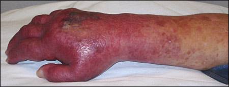
The discoloration and edema extended proximally over the next several days with the formation of a bullous lesion on the dorsum of her hand (Figure 1). She remained afebrile, her white blood cell count was normal, and she denied feeling pain, although her previous stroke had left her with little sensory or motor function on that side. Her overall clinical condition improved steadily.
Figure 1.
Her medications from home included propranolol (Inderal), phenytoin, warfarin (Coumadin), fluvastatin (Lescol), and escitalopram (Lexapro). She received no medications to which she had a known allergy. Her previous surgeries included bilateral mastectomy and a left femoral thrombectomy.
Question
Based on the patient's history and physical examination, which of the following is the most likely cause of the edema and discoloration?
A. Adverse reaction to intravenous phenytoin.
B. Cellulitis secondary to intravenous site infection.
C. Necrotizing fasciitis.
D. Normal saline intravenous fluid infiltration.
E. Subclavian vein thrombosis.
Discussion
The answer is A: adverse reaction to intravenous phenytoin. Phenytoin has been used to treat acute seizures since the 1950s. Even appropriate use of intravenous phenytoin has led to what is known as “purple glove” syndrome. Presentation generally includes progressive distal limb edema, discoloration, and pain soon after administration.1 Skin necrosis, limb ischemia, vascular compression, and compartment syndrome may occur. Fasciotomy, skin grafting, and even limb amputation may be required.1,2
Typically, within two to 12 hours after infusion, erythema and blue-purple discoloration occur around the intravenous site, and there may be petechiae on the fingers and palms. Then, 12 to 24 hours after infusion, spreading discoloration, edema, skin blistering, sloughing, and ulcerations may occur, with possible extension thereafter. Resolution may take weeks to months, with discoloration receding toward the original intravenous site.1,3 Incidence has been reported as 5.9 percent.1
There are several possible explanations for the development of purple glove syndrome. First, sodium hydroxide, propylene glycol, and ethanol are added to the phenytoin solution to raise the pH level to 12 and enhance solubility. This highly alkaline solution may induce vasoconstriction and thrombosis in vessels, encouraging leakage into interstitial spaces. A second explanation is that the mixing of the alkaline solution with blood may induce precipitation of phenytoin crystals, leading to vessel obstruction and leakage. Another possibility is that the alkaline solution may break down endothelial intercellular junctions and allow leakage. In some cases, simple infiltration occurs after defects in the vessel walls are caused by intravenous cannulation. Usually, there is no obvious sign of extravasation during administration of intravenous phenytoin.1 Although ultrasonography showed no deep venous thrombosis in this patient, it did reveal superficial thrombophlebitis of the entire length of her cephalic vein.
The medical literature recommends limb elevation and gentle, dry heat in affected patients. Frequent assessment of pulses, capillary refill, edema, and discoloration is essential.3 Consultation with a vascular surgeon is prudent even in cases that seem to be mild. Pain is often intense and may require narcotics. It has been suggested that pain out of proportion to the use of an intravenous catheter might be an early sign of purple glove syndrome. Unfortunately, many patients who receive intravenous phenytoin are unable to communicate. Use of the affected limb may be limited for some time as recovery proceeds.
Risk factors for purple glove syndrome may include multiple intravenous doses, higher rates of intravenous administration (even if lower than the labeling specifies), age older than 60 years, vascular disease, higher initial and 24-hour total intravenous doses, use of angiocatheters smaller than 20 g, flush solutions containing benzyl alcohol, use in the acute setting, and female sex.1–3
It has been suggested that the more costly prodrug fosphenytoin (Cerebyx), which is highly water soluble at neutral pH levels and is converted to phenytoin in the body, be given to prevent purple glove syndrome and other adverse effects. It can be administered intravenously (or even intramuscularly, unlike phenytoin) at rates higher than phenytoin. It is purported to be much less painful if extravasation does occur.1
Cellulitis often presents as an erythematous, edematous, irregularly shaped plaque, possibly following a skin injury. The mid to deep dermis is infected, often with Streptococcus, Staphylococcus, or Pseudomonas spp. (especially in immunocompromised patients who may also have fungal cellulitis). Pasteurella multocida occurs after animal bites. Cellulitis may be accompanied by leukocytosis, fever, malaise, and lymph-adenopathy. Appropriate antibiotics must be given; surgical debridement is sometimes necessary.4
Necrotizing fasciitis is a fairly rare, deeper infection occurring between the fascial planes, often following a skin injury. Affected patients may present with limb edema, dusky red-purple discoloration, bullae, and severe pain that may progress to anesthesia. It can extend rapidly between fascial planes; cause crepitus because of gas production from various organisms; and may be multiorganismal, including aerobes and anaerobes. It can cause severe tissue necrosis and gangrene, and can lead to extensive systemic effects such as tachycardia, hypotension, fever, and even death. It must be treated with early, aggressive, and repeated surgical debridement; powerful antibiotics; and supportive care.4
Discomfort and localized edema often accompany intravenous infiltration. At least initially, there is no discoloration of the skin if the infiltrated solution is innocuous, such as normal saline. Treatment involves discontinuation of fluids in that area, limb elevation, and time.
Subclavian vein thrombosis may present with limb edema, cyanosis of the hand or fingers on the affected side, and dilated subcutaneous collateral veins in the upper arm and chest. The diagnosis is generally confirmed via ultrasonography. An underlying hyper-coagulable state should be investigated when appropriate. Anomalous anatomic structures may require surgical intervention. Long-term indwelling catheters can be causative. Treatment of subclavian vein thrombosis may include thrombolysis or thrombectomy.5
Selected Differential Diagnosis of Distal Upper Extremity Edema and Discoloration
| Condition | Characteristics |
|---|---|
| Adverse reaction to IV phenytoin | Progressive distal limb edema; blue-purple discoloration; severe pain; worsens even after IV phenytoin stopped |
| Cellulitis secondary to IV site infection | Slow onset; skin is red and hot; purulent discharge possible; usually lacks blue-purple discoloration; plaque is shiny, very tender, with irregular border |
| Necrotizing fasciitis | Tissue necrosis; putrid discharge, bullae, gas production; extension via rapid burrowing through fascial planes; may have dusky red-purple discoloration |
| Normal saline IV fluid infiltration | Noticeable, often pitting, edema; may become erythematous but lacks blue-purple discoloration |
| Subclavian vein thrombosis | Mild-to-moderate nonpitting edema and mild cyanosis of the hand and fingers on the affected side; dilated subcutaneous collateral veins may be present over the upper arm and chest |
IV = intravenous.
