Am Fam Physician. 2007;75(9):1379-1380
Author disclosure: Nothing to disclose.
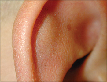
A 22-year-old man presented for evaluation of a painless swelling on his right ear that he first noticed about five months earlier. He denied any history of trauma. The patient was otherwise healthy with no significant medical history. The scaphoid fossa of the right auricle demonstrated a nontender, noninflammatory, almond-shaped nodule approximately 1.5 cm × 1 cm (see accompanying figure).

Question
Discussion
The answer is C: pseudocyst of the auricle. An auricular pseudocyst, also referred to as benign idiopathic cystic chondromalacia, is an uncommon condition characterized by a painless, cystic swelling of the concha of the pinna.1 It most commonly affects healthy young men2 and rarely is there a history of antecedent trauma. There appears to be no racial predisposition.3 The exact etiology is unknown, but it is thought to be related to degenerative cartilage.4
The diagnosis can usually be made on clinical grounds, and routine histology is rarely necessary. However, when biopsy is performed, a characteristic finding is an intracartilaginous space with no epithelial lining.3 Needle aspiration of the cyst typically reveals a clear or yellow sterile fluid with the consistency of olive oil.5
Treatment goals include permanent removal of the pseudocyst with good aesthetic outcome and preservation of the normal anatomic architecture.3 It should be noted that simple needle aspiration of the pseudocyst, without additional treatment, always results in prompt reaccumulation of fluid, usually in less than three days.3 Definitive treatment by surgical excision with pressure dressing or compression buttoning usually results in a satisfying outcome; surgical excision remains the treatment of choice.6
Other treatment options include high-dose oral corticosteroids, simple aspiration with compression bolsters, and punch biopsy with pressure dressing.7 Intralesional injection of corticosteroids,8,9 sclerosing agents,10 and minocycline4 also have been attempted with varying degrees of success. Aspiration followed by intralesional triamcinolone acetonide (Kenalog) is another option and may be attempted up to three times before moving to surgical treatment.8,9
Chondrodermatitis nodularis chronica helicis, the most common diagnosis to exclude in the work-up, typically presents as an exquisitely tender nodule on the outer helix that may interfere with sleep. There is often a preceding history of trauma. It is most common in men older than 40 years.
An othematoma invariably involves a history of notable trauma and may demonstrate clinical signs of inflammation, including pain and redness.
Relapsing polychondritis is a rare multisystem autoimmune disease that can affect all cartilage throughout the body, including the ear, eyes, nose, and respiratory system.11 Relapsing polychondritis of the ear is manifested as a painful, red, swollen superior portion of the pinna, sparing the noncartilaginous ear lobe.
Subperichondral abscess typically presents with pain and surrounding erythema. Diagnosis can be confirmed by aspiration of purulent fluid.
| Condition | Characteristics |
|---|---|
| Chondrodermatitis nodularis chronica helicis | Exquisitely tender nodule on the outer helix; often a history of trauma |
| Othematoma | History of trauma; may demonstrate pain and redness |
| Pseudocyst of the auricle | Painless, cystic swelling of the concha of the pinna; usually no history of trauma |
| Relapsing polychondritis | Autoimmune cartilage disease; ear manifestations are a painful, red, swollen superior portion of the pinna, sparing the noncartilaginous earlobe |
| Subperichondral abscess | Pain and surrounding erythema; purulent aspirate |