A 19-month-old, previously healthy Hispanic child was found to have anemia during a routine screening examination; her hemoglobin level was 5.4 g per dL (54 g per L). The child had a recent history of constipation that lasted two months and was being treated with lactulose (Constulose). The parents denied a history of irritability, lethargy, or abdominal pain in the child. The family lived in an older neighborhood and drank tap water from the city's water supply.
The child's physical examination findings were unremarkable, except for notable pallor. She had normal development, was attentive, and did not appear to be distressed. Laboratory testing revealed a mean corpuscular volume of 49 μm3 (49 fL) and a white blood cell count of 2,580 per mm3 (2.6 × 10⁹ per L) with 66 percent lymphocytes, 20 percent neutrophils, and 2 percent bands. The peripheral blood smear showed round, dark-blue granules within the red blood cells. During the workup, radiography of the child's abdomen and long bones was performed (Figures 1 and2).
Figure 1.
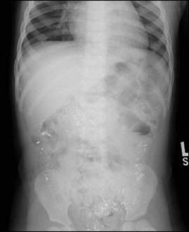
Radiograph of the abdomen in a 19-month-old child.
Figure 2.
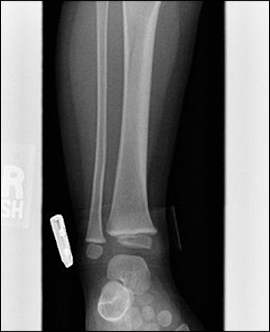
Radiograph of the forearm in a 19-month-old child.
Question
Based on the patient's history and physical examination, which one of the following is the most likely diagnosis?
A. Beta-thalassemia major.
B. Hirschsprung's disease.
C. Lead toxicity.
D. Sickle cell anemia.
E. Sideroblastic anemia.
Discussion
The answer is C: lead toxicity. The patient's blood lead level was confirmed to be 70 mcg per dL (3.38 μmol per L). Lead is absorbed primarily by ingesting or inhaling inorganic dust particles. Children are at the highest risk of toxicity because they absorb proportionately larger amounts of lead during an exposure.
Furthermore, children younger than four years are more susceptible to the toxic effects of lead because they have an incomplete blood-brain barrier, a predisposition to iron deficiency anemia, and increased exposure from crawling on the floor and hand-to-mouth activity. Lead causes anemia by directly inhibiting δ-aminolevulinate synthase and dehydratase, enzymes necessary for heme biosynthesis. Lead also inhibits the enzyme ferrochelatase, which leads to increased zinc protoporphyrin levels in the blood.1
The Centers for Disease Control and Prevention defines lead toxicity as a blood lead level of 10 mcg per dL (0.48 μmol per L) or more.2 A normal blood lead level is 0.
Although most children with elevated blood lead levels are asymptomatic at the time of screening, marked neurocognitive defects have been demonstrated even in children with levels of less than 10 mcg per dL.3 Effects of lead poisoning include irritability, behavior changes, altered sleep, nephropathy, colic, constipation, delayed bone development, and gingival lead lines. Severe toxicity (i.e., blood lead levels of more than 70 mcg per dL) may cause encephalopathy, paralysis, seizures, or death.4
Radiography may show abdominal lead flecks (Figure 1) and “lead lines” at the end of growing long bones (Figure 2).5 The peripheral blood smear may show basophilic stippling (as described in our patient).
Treatment of lead poisoning includes environmental avoidance, appropriate nutrition, chelation therapy (depending on blood lead level), and supportive care. However, primary prevention remains the best option.
Beta-thalassemia major is the most serious type of thalassemia. It typically presents between six and 12 months of age as profound anemia, massive hepato-splenomegaly, growth delay, jaundice, and bone deformities. The peripheral blood smear may reveal microcytic anemia with severe hypochromia, anisocytosis, and fragmented and nucleated red blood cells.
In patients with Hirschsprung's disease, the absence of autonomic plexuses in the colonic wall causes functional obstruction and constipation. The condition is usually diagnosed in newborns or within a few months of birth. However, less severe disease may be diagnosed in adulthood in patients with chronic constipation and failure to thrive. Severe anemia is unlikely unless there is bleeding from surgery or total colonic aganglionosis.6
Sickle cell anemia is an inherited hemoglobin disorder. In the United States, about 8 percent of black persons carry the gene.7 The primary mechanism of anemia is extravascular hemolysis. The peripheral blood smear may show characteristic sickle-shaped red blood cells.
Sideroblastic anemia may be inherited or acquired and is caused by defects in heme synthesis, which leads to iron retention within the mitochondria. Although it is usually diagnosed in adults, hereditary sideroblastic anemia also may occur in children. Clinical findings may include hepatosplenomegaly, jaundice, pallor, and elevated serum ferritin and iron levels.
Selected Differential Diagnosis of Anemia in a Toddler
| Condition | Characteristics |
|---|---|
| Beta-thalassemia major | Typically presents between six and 12 months of age; ineffective erythropoiesis; high mortality during first five years of life |
| Chronic disease | Usually normocytic, although it may be microcytic; anemia may be associated with normal or elevated serum ferritin level, low serum iron level, and low total iron-binding capacity |
| Dietary iron deficiency | Low serum iron and ferritin levels with high total iron-binding capacity; associated with exclusive consumption of cow's milk |
| Lead toxicity | History of pica and living in older homes; blood lead levels are diagnostic |
| Sideroblastic anemia | Hereditary type occurs in children; peripheral blood smear shows ringed sideroblasts; increased serum iron, ferritin, and transferrin saturation; decreased transferrin saturation with increased bone marrow iron |
