
Am Fam Physician. 2008;77(8):1151-1152
Author disclosure: Nothing to disclose.
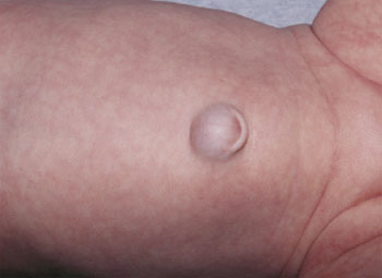
A six-month-old girl presented for a well-child visit. On abdominal examination, a periumbilical mass, 1 cm in diameter, was noted (see accompanying figure). The mass was soft and reduced completely on palpation with no apparent pain. A circular defect, 0.5 cm in diameter, that was distinct and superior to the umbilical scar was visible in the underlying fascia. The swelling became more prominent when the infant cried.
The mass had been present for a few months and had gradually increased in size. The infant was born at term and had umbilical cord separation at seven days of age; there was no noticeable umbilical swelling at birth. She had no history of pain, discharge, gastrointestinal problems, abdominal surgery, or local trauma.
Question
Discussion
The answer is A: epigastric hernia. An epigastric hernia is caused by a defect in the linea alba between the xiphoid process and the umbilicus, and it usually occurs just superior to the umbilicus. Epigastric hernias occur in 3 to 5 percent of the population and are most likely to affect men 20 to 50 years of age. Among those with the condition, 20 to 30 percent have multiple defects.1 On examination, an epigastric hernia tends to push the umbilicus downward into a U shape (see accompanying figure). Most patients are asymptomatic, but the hernia may become incarcerated and painful. Ultrasonography or computed tomography may help confirm the diagnosis. Surgical repair is needed because of the risk of painful strangulation; these defects do not close spontaneously.
Gastroschisis and omphalocele are uncommon congenital hernias caused by abnormal abdominal wall development. The incidence of gastroschisis is 1.0 in 10,000, and the incidence of omphalocele is 2.5 in 10,000.4 Omphalocele is characterized by extension of abdominal viscera, often including the liver, into the umbilical stalk. The exposed viscera are covered by a translucent, bilaminar sac. Associated genetic abnormalities, such as trisomy 18 syndrome, are often present.
An umbilical granuloma is a red, solid, velvety mass on the umbilicus occurring in the first few weeks of life. Serous or serosanguineous discharge may occur. An umbilical granuloma may be treated with chemical cautery using silver nitrate. Although surgery is rarely needed, a granuloma that persists despite repeated treatment warrants further evaluation to rule out other pathology.5,6
Umbilical hernias in children occur in the first few months of life 1 and are caused by incomplete closure of the umbilical ring. They occur more often in black girls, premature infants, and children with Down syndrome or hypothyroidism.7 The hernia appears as a skin-covered swelling, which pushes the umbilicus outward, and becomes more prominent when the child cries or strains. In contrast to epigastric hernias, most childhood umbilical hernias close spontaneously by four or five years of age. However, spontaneous closure is less likely when the hernia is larger than 2 cm in diameter. Umbilical hernias in children rarely become incarcerated (one in 1,500 cases).8 Surgical repair is needed in children older than four years, for very large hernias (greater than 2 cm in diameter), and for incarcerated hernias.1
| Condition | Characteristics |
|---|---|
| Epigastric hernia | Small, midline defect in the linea alba leading to a hernia; on palpation, the defect can be differentiated from the intact umbilical ring |
| Gastroschisis | Exposed extracorporeal viscera not covered by a sac; herniates lateral to the intact umbilicus |
| Omphalocele | Extension of abdominal viscera covered by a translucent, bilaminar sac; herniates through the umbilicus; umbilical cord attaches to the sac |
| Umbilical granuloma | Red, moist, small, solid mass on the umbilicus |
| Umbilical hernia | Skin-covered, reducible hernia caused by incomplete closure of the umbilical ring |