A six-month-old girl presented with brownish macules all over her body (Figures 1 and 2). The child's mother reported that the macules, which appeared at birth, became red and swollen when scratched or rubbed, and decreased in size as the child grew. The pregnancy and birth were normal, and the child had no family history of skin lesions or cancers. The child's workup at birth was unremarkable, and her development was progressing normally.
Figure 1.
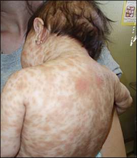
Figure 2.
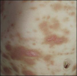
On physical examination, the patient was active, responded appropriately to visual and auditory stimuli, and had normal vital signs. The oval to round skin lesions were approximately 0.5 to 1.5 cm in diameter, had well-demarcated borders, and involved all skin areas except the soles, palms, and mucous membranes.
Question
Based on the patient's history and physical examination, which one of the following is the most likely diagnosis?
A. Neurofibromatosis 1.
B. Peutz-Jeghers syndrome.
C. Urticaria pigmentosa.
D. Xeroderma pigmentosum.
Discussion
The answer is C: urticaria pigmentosa. Urticaria pigmentosa is a type of cutaneous mastocytosis characterized by aggregates of mast cells in the dermis, leading to the development of dark yellow to brown macules.1 Mastocytosis encompasses a spectrum of disorders that range from a solitary cutaneous nodule to diffuse infiltration of skin with involvement of other organs.2 Urticaria pigmentosa is a common type of mastocytosis in children and is considered a hyperplastic, rather than a neoplastic, disorder.1,2 There are two types of urticaria pigmentosa. In the classic type (infantile onset), lesions are present at birth or erupt during the first two years of life.1 Lesions may be macular, nodular, papular, vesicular, or bullous and often have a symmetric distribution.1 The palms and soles are spared.2 Rubbing the lesions usually produces erythema and whealing (Darier sign). Dermographism of normal skin is often present.1 Histopathology of the skin lesions confirms the diagnosis.2 The nonclassic type (adult onset) is caused by a mutation of a stem cell factor.1 The lesions are similar to the classic type and may develop at any time from infancy to adulthood. The lesions do not resolve, new lesions continually develop, and systemic involvement is more common.1
Clinical manifestations of mastocytosis depend on the organ or systems affected. Cutaneous manifestations may include pruritus, episodic flushing, and the characteristic rash.1 Pruritus may be exacerbated by changes in climate, skin friction, spicy foods, ethanol, exercise, stinging insects, radiocontrast media, infections, and use of aspirin or other medications.3 Patients with the classic type have a good prognosis, often with spontaneous resolution of lesions by four years of age.1
A bone marrow biopsy should be performed in patients with urticaria pigmentosa if peripheral blood findings are abnormal or if the nonclassic type is suspected.3
Neurofibromatosis 1 is an autosomal dominant disease affecting neural crest–derived cells. It is characterized by café au lait spots and other light-colored to brown macules, and is associated with multiple neural tumors.4 The macules may be present at birth, and usually increase in size and number with age.
Peutz-Jeghers syndrome is an autosomal dominant disease characterized by melanotic macules on the lips and mucous membranes with gastric polyposis.1 The pigmented macules, which begin to appear in infancy, can be up to 2 cm in size.1 Lesions may develop on the hands, feet, nose, and around the eyes and umbilicus.1
Xeroderma pigmentosum is an autosomal recessive disease affecting sun-exposed skin, such as on the face, arms, and legs. Skin changes may occur in infancy and include reddening with scaling; freckling; and irregular dark spots, which may be premalignant.1
Selected Differential Diagnosis of Brown Macules in Infancy
| Condition | Characteristics |
|---|---|
| Neurofibromatosis 1 | Café au lait spots or other light-colored to brown macules; associated with multiple neural tumors; macules usually increase in size and number with age |
| Peutz-Jeghers syndrome | Melanotic macules, usually on the lips and mucous membranes, with gastrointestinal polyposis; macules may develop on the hands, feet, nose, and around the eyes and umbilicus; macules are up to 2 cm in size |
| Urticaria pigmentosa | Macular, nodular, or popular lesions ranging in color from dark yellow to brown; often symmetrically distributed; erythema and whealing (Darier sign); dermographism of normal skin is often present |
| Xeroderma pigmentosum | Affects sun-exposed skin, such as on the face, arms, and legs; reddening of the skin with scaling; freckling; irregular dark spots, which may be premalignant |
