Although eye pain is often accompanied by redness or injection, pain can also occur with a quiet eye. Pain in a quiet eye can be the first sign of a vision-threatening condition, a more benign ophthalmologic condition, or a nonophthalmologic condition. Acute narrow-angle glaucoma is an emergent vision-threatening condition that requires immediate treatment and referral to an ophthalmologist. Although most nonophthalmologic conditions that cause eye pain do not need immediate treatment, giant cell (temporal) arteritis requires urgent treatment with corticosteroids. Other vascular conditions, such as carotid artery disease, thrombosis of the cavernous sinus, and transient ischemic attack or stroke, rarely cause eye pain but must be considered. Pain may also be referred from the sinuses or from neurologic conditions, such as trigeminal neuralgia, migraine and cluster headaches, and increased intracranial pressure. The differential diagnosis of eye pain in the quiet eye is extensive, necessitating a systematic and thorough approach.
Pain in an eye without redness or injection (quiet eye) is uncommon but may represent a serious condition. There are five anatomic areas to consider when determining etiology: ocular, orbital, cranial, neurologic, and vascular. The first step is determining if the pain is truly coming from the eye and if it is caused by a vision-threatening condition.
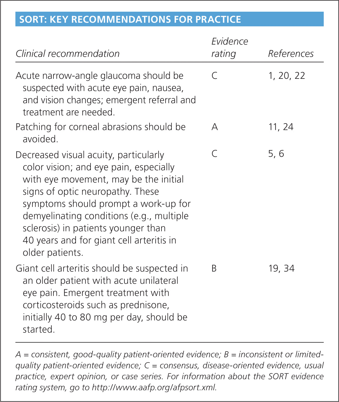
SORT: KEY RECOMMENDATIONS FOR PRACTICE
| Clinical recommendation | Evidence rating | References |
|---|---|---|
| Acute narrow-angle glaucoma should be suspected with acute eye pain, nausea, and vision changes; emergent referral and treatment are needed. | C | 1, 20, 22 |
| Patching for corneal abrasions should be avoided. | A | 11, 24 |
| Decreased visual acuity, particularly color vision; and eye pain, especially with eye movement, may be the initial signs of optic neuropathy. These symptoms should prompt a work-up for demyelinating conditions (e.g., multiple sclerosis) in patients younger than 40 years and for giant cell arteritis in older patients. | C | 5, 6 |
| Giant cell arteritis should be suspected in an older patient with acute unilateral eye pain. Emergent treatment with corticosteroids such as prednisone, initially 40 to 80 mg per day, should be started. | B | 19, 34 |
A = consistent, good-quality patient-oriented evidence; B = inconsistent or limited-quality patient-oriented evidence; C = consensus, disease-oriented evidence, usual practice, expert opinion, or case series. For information about the SORT evidence rating system, go to https://www.aafp.org/afpsort.xml.
Evaluation
History and examination findings may point to a general etiology or specific condition (Table 1).1–10 If the history suggests a systemic or neurologic condition and examination confirms no visual impairment, a vision-threatening lesion is much less likely. A history of similar pain that may have resolved spontaneously could suggest neurologic causes, such as trigeminal neuralgia, migraine and cluster headaches, and transient ischemic attack. Palpation of the orbits and measurement of intraocular pressure can rule out narrow-angle glaucoma. Palpation of the temporal arteries should be performed next, because temporal arteritis is a relatively common cause of vision-threatening eye pain. Figure 1 is an algorithm for the evaluation and treatment of pain in the quiet eye.1–3,11–19
Table 1. History and Physical Examination Findings Suggestive of Causes of Pain in the Quiet, Nonred Eye
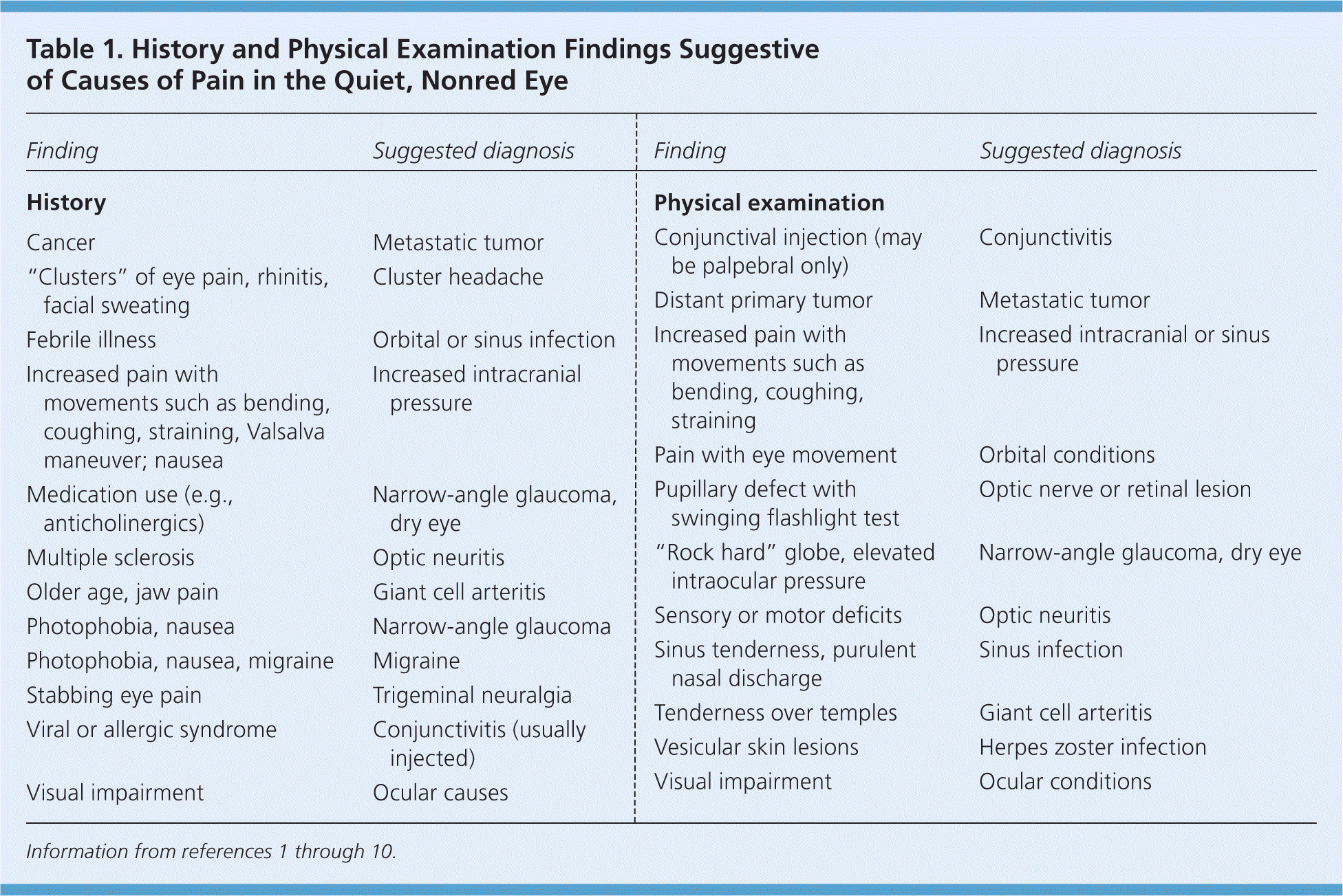
| Finding | Suggested diagnosis |
|---|---|
| History | |
| Cancer | Metastatic tumor |
| “Clusters” of eye pain, rhinitis, facial sweating | Cluster headache |
| Febrile illness | Orbital or sinus infection |
| Increased pain with movements such as bending, coughing, straining, Valsalva maneuver; nausea | Increased intracranial pressure |
| Medication use (e.g., anticholinergics) | Narrow-angle glaucoma, dry eye |
| Multiple sclerosis | Optic neuritis |
| Older age, jaw pain | Giant cell arteritis |
| Photophobia, nausea | Narrow-angle glaucoma |
| Photophobia, nausea, migraine | Migraine |
| Stabbing eye pain | Trigeminal neuralgia |
| Viral or allergic syndrome | Conjunctivitis (usually injected) |
| Visual impairment | Ocular causes |
| Physical examination | |
| Conjunctival injection (may be palpebral only) | Conjunctivitis |
| Distant primary tumor | Metastatic tumor |
| Increased pain with movements such as bending, coughing, straining | Increased intracranial or sinus pressure |
| Pain with eye movement | Orbital conditions |
| Pupillary defect with swinging flashlight test | Optic nerve or retinal lesion |
| “Rock hard” globe, elevated intraocular pressure | Narrow-angle glaucoma, dry eye |
| Sensory or motor deficits | Optic neuritis |
| Sinus tenderness, purulent nasal discharge | Sinus infection |
| Tenderness over temples | Giant cell arteritis |
| Vesicular skin lesions | Herpes zoster infection |
| Visual impairment | Ocular conditions |
Figure 1. Evaluation and Treatment of Pain in the Quiet, Nonred Eye
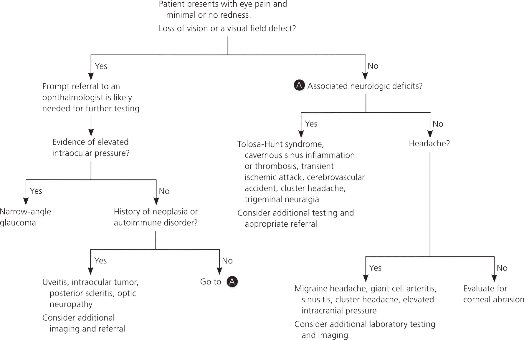
Algorithm for the evaluation and treatment of pain in the quiet, nonred eye.
Information from references 1 through 3, and 11 through 19.
Ocular Conditions
NARROW-ANGLE GLAUCOMA
Narrow-angle glaucoma is one of the most common vision-threatening causes of acute pain in the quiet eye. The annual incidence of narrow-angle glaucoma ranges from one to 30 per 1,000 persons, depending on age and ethnicity.1,20,21 Persons with narrow-angle glaucoma classically present with headache or eye pain, vision changes, and nausea and vomiting. Although the eye is usually inflamed, inflammation may be absent. In persons with a shallow anterior chamber, the sudden increase in intraocular pressure may be precipitated by anything that dilates the pupil, such as anticholinergics, adrenergic stimulation, or dim lighting.22 Although classic narrow-angle glaucoma is easy to identify, the variability in presentation and the possible absence or minimization of visual changes can make diagnosis difficult. Patients often describe having a visual halo, and subtle conjunctival injection or corneal edema may be noted on examination.2 The affected pupil may be unreactive and mildly dilated. Referral to an ophthalmologist for definitive evaluation and treatment is warranted.
CORNEAL DISEASES
The most common corneal causes of eye pain are corneal infections, abrasions, and foreign bodies. These conditions are typically associated with redness and injection. Patients with small foreign bodies or abrasions, however, may initially present with eye pain and only minimal injection.1,3 Keratitis (inflammation of the cornea) may produce severe pain due to the rich innervation of the cornea. Viral infections (e.g., herpes simplex virus), bacterial infections, exposure to ultraviolet light or chemicals, and inflammatory conditions can all cause keratitis.1 Treatment of corneal abrasions typically includes oral analgesics and topical antibiotics, although corneal patching should be avoided.3,11,23,24 If keratitis is suspected, prompt referral to an ophthalmologist is warranted.
INTRAOCULAR TUMORS
Primary intraocular tumors (choroidal melanoma) and metastatic tumors rarely cause eye pain. However, orbital extension of the tumor may involve the trigeminal nerve, leading to pain. Intraocular tumors may also cause inflammatory reactions or raised intraorbital pressure, both of which can cause pain.2 The most common primary cancers leading to intraocular metastases are breast, lung, and gastrointestinal cancers.4,25
OPTIC NEUROPATHY
Optic neuropathy typically presents as decreased visual acuity, particularly color vision; and eye pain, especially with eye movement.5,6 In patients younger than 40 years, demyelinating conditions (e.g., multiple sclerosis) are the most common causes of optic neuritis, and urgent referral to a neurologist and ophthalmologist is necessary.1,2,6 Because older patients presenting with possible optic neuritis are more likely to have giant cell (temporal) arteritis, an erythrocyte sedimentation rate or C-reactive protein level should be obtained. If optic neuropathy is clinically suspected, prompt corticosteroid therapy and referral are indicated while awaiting test results.2
DRY EYE
Dry eye may lead to eye pain, foreign body sensation, tearing, photophobia, and occasional redness. Causes include contact use; medications (e.g., antihistamines, clonidine [Catapres], beta blockers, tricyclic antidepressants, isotretinoin [formerly Accutane], angiotensin-converting enzyme inhibitors, opiates, scopolamine); systemic diseases (e.g., keratoconjunctivitis sicca, Sjögren syndrome, rheumatoid arthritis); trauma; and environmental factors (e.g., air conditioning).12
Orbital Conditions
Orbital inflammation, infection, and tumor invasion can lead to eye pain. Findings suggestive of an orbital cause of eye pain include optic neuropathy, limitation or pain with ocular movement, diplopia, proptosis, enophthalmos, gaze evoked amaurosis or facial pain, and paresthesia.1,2 Orbital vascular lesions may produce symptoms intermittently with movements such as coughing, straining, or bending. If an orbital lesion is suspected, imaging with ultrasonography, computed tomography, or magnetic resonance imaging and referral to an ophthalmologist are recommended.1,2
Cranial Conditions
Cavernous sinus thrombosis is caused by spreading infection and may lead to eye pain via irritation of the third cranial nerve, other oculomotor nerves, or occasionally the ophthalmic branch of the trigeminal nerve.2 Cavernous sinus thrombosis should be suspected with rapid onset of unilateral periorbital swelling, photophobia, chemosis, proptosis, headache, or cranial nerve palsies. The condition typically spreads to the contralateral eye within 48 hours.7
Tolosa-Hunt syndrome is an idiopathic inflammatory, granulomatous condition of the cavernous sinus that causes eye and facial pain. Key to diagnosis is resolution within 72 hours of starting corticosteroid therapy.13
Paranasal sinusitis may also cause pain in the quiet eye. Pain from frontal sinus disease classically extends diffusely over the forehead, maxillary sinus pain extends to the cheek and lower orbit, and ethmoid and sphenoid sinusitis pain extends to the orbits or vertex of the skull. Sphenoid sinusitis should prompt referral to an otolaryngologist or neurosurgeon because it may be associated with serious neurologic conditions.26
Neurologic Conditions
CLUSTER HEADACHE
Cluster headaches are most common in younger men and produce severe unilateral eye pain. Typically, the pain lasts from 15 to 45 minutes, but can persist for up to three hours. The pain is debilitating and is usually located in or directly behind the eye. The headaches tend to come in clusters with multiple attacks occurring throughout the day.14,27
Patients with cluster headache may also have manifestations of Horner syndrome (e.g., ptosis, miosis, anisocoria), rhinorrhea, nasal congestion, and facial sweating. The condition is more common in patients who use tobacco and alcohol, and may be triggered by nitrates. Sumatriptan injections (Imitrex), intranasal triptans, and high-dose oral triptans appear to be the most effective treatments.15,28 Treatment with high-flow oxygen is common, although there is minimal evidence to support its use.29
MIGRAINE HEADACHE
Migraine headaches are rarely associated with isolated eye pain, although migraine pain may radiate to the eye. Patients with migraine may have other ocular symptoms, including scotomas, photophobia, blurred vision, vision loss, ptosis, lacrimation, and diplopia.8 Ocular migraines and migraines with pain around the eye should be treated as typical migraines.30
TRIGEMINAL NEURALGIA
Trigeminal neuralgia typically produces episodic, severe, unilateral facial pain accompanied by numbness and sensory loss.2,9,16,31 Because the ophthalmic division of the trigeminal nerve provides sensation to the eye, the condition may also cause eye pain. The pain tends to recur and worsens with time.9,16
ELEVATED INTRACRANIAL PRESSURE
Any condition that causes elevated intracranial pressure, such as cerebral aneurysm, brain tumors, other lesions, venous sinus thrombosis, and pseudotumor cerebri, can also lead to eye pain and headache. Eye pain caused by elevated intracranial pressure often can be exacerbated by Valsalva maneuver.32
Vascular Conditions
Rarely, eye pain is the presenting symptom of subdural, epidural, subarachnoid, or intracerebral hemorrhage and is caused by pain receptors within the arteries that are innervated by the ophthalmic branch of the trigeminal nerve. Among patients with a stroke in the internal or middle cerebral artery distribution, 25 percent have pain in the orbit or frontal region ipsilateral to the infarction.33
Any carotid artery disease (e.g., inflammation, emboli, thrombosis, dissection) can lead to monocular symptoms and may herald an impending stroke, although isolated ocular pain would not be the only symptom. Horner syndrome on the side of the arterial injury may also occur.17,18
Giant cell arteritis typically manifests as tenderness over the temple in patients older than 50 years, and is often accompanied by jaw claudication and diplopia.34 Patients may also have associated eye pain and visual loss or blurred vision as a result of orbital ischemia. An elevated erythrocyte sedimentation rate (greater than 50 mm per hour) or C-reactive protein level increases the likelihood of giant cell arteritis. Emergent treatment, before diagnosis is confirmed, consists of high-dose steroids such as prednisone, initially 40 to 80 mg per day.19,34 If the condition is untreated, vision loss can occur from involvement of the ophthalmic artery.10,19
