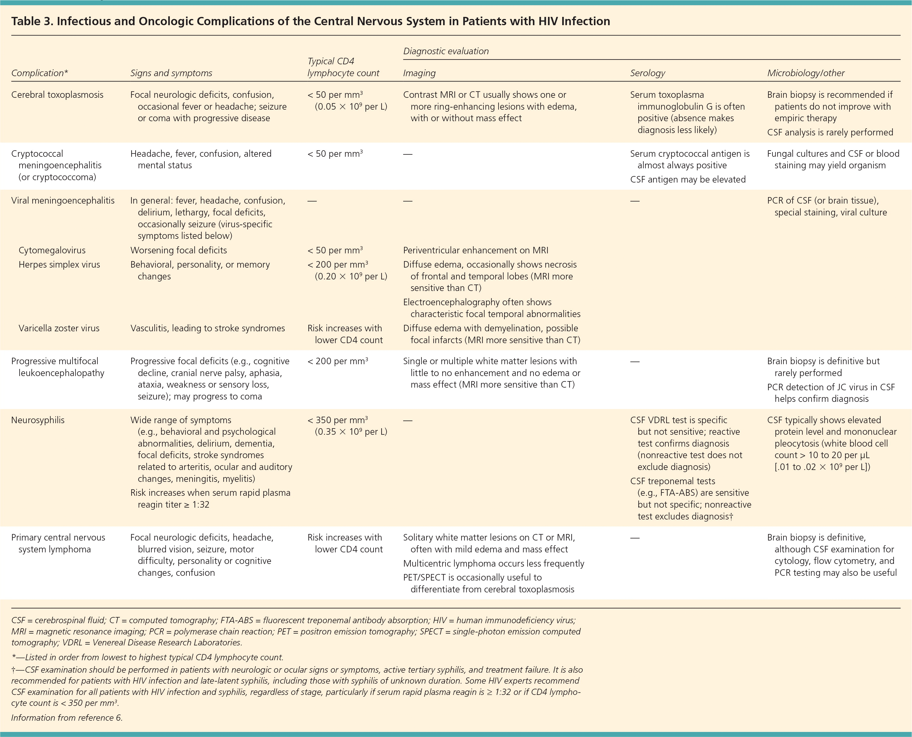
| Complication* | Signs and symptoms | Typical CD4 lymphocyte count | Diagnostic evaluation | |||
|---|---|---|---|---|---|---|
| Imaging | Serology | Microbiology/other | ||||
| Cerebral toxoplasmosis | Focal neurologic deficits, confusion, occasional fever or headache; seizure or coma with progressive disease | < 50 per mm3 (0.05 × 10⁹ per L) | Contrast MRI or CT usually shows one or more ring-enhancing lesions with edema, with or without mass effect | Serum toxoplasma immunoglobulin G is often positive (absence makes diagnosis less likely) | Brain biopsy is recommended if patients do not improve with empiric therapy | |
| CSF analysis is rarely performed | ||||||
| Cryptococcal meningoencephalitis (or cryptococcoma) | Headache, fever, confusion, altered mental status | < 50 per mm3 | — | Serum cryptococcal antigen is almost always positive | Fungal cultures and CSF or blood staining may yield organism | |
| CSF antigen may be elevated | ||||||
| Viral meningoencephalitis | In general: fever, headache, confusion, delirium, lethargy, focal deficits, occasionally seizure (virus-specific symptoms listed below) | — | — | — | PCR of CSF (or brain tissue), special staining, viral culture | |
| Cytomegalovirus | Worsening focal deficits | < 50 per mm3 | Periventricular enhancement on MRI | |||
| Herpes simplex virus | Behavioral, personality, or memory changes | < 200 per mm3 (0.20 × 10⁹ per L) | Diffuse edema, occasionally shows necrosis of frontal and temporal lobes (MRI more sensitive than CT) | |||
| Electroencephalography often shows characteristic focal temporal abnormalities | ||||||
| Varicella zoster virus | Vasculitis, leading to stroke syndromes | Risk increases with lower CD4 count | Diffuse edema with demyelination, possible focal infarcts (MRI more sensitive than CT) | |||
| Progressive multifocal leukoencephalopathy | Progressive focal deficits (e.g., cognitive decline, cranial nerve palsy, aphasia, ataxia, weakness or sensory loss, seizure); may progress to coma | < 200 per mm3 | Single or multiple white matter lesions with little to no enhancement and no edema or mass effect (MRI more sensitive than CT) | — | Brain biopsy is definitive but rarely performed | |
| PCR detection of JC virus in CSF helps confirm diagnosis | ||||||
| Neurosyphilis | Wide range of symptoms (e.g., behavioral and psychological abnormalities, delirium, dementia, focal deficits, stroke syndromes related to arteritis, ocular and auditory changes, meningitis, myelitis) | < 350 per mm3 (0.35 × 10⁹ per L) | — | CSF VDRL test is specific but not sensitive; reactive test confirms diagnosis (nonreactive test does not exclude diagnosis) CSF treponemal tests (e.g., FTA-ABS) are sensitive but not specific; nonreactive test excludes diagnosis† | CSF typically shows elevated protein level and mononuclear pleocytosis (white blood cell count > 10 to 20 per μL [.01 to .02 × 109 per L]) | |
| Risk increases when serum rapid plasma reagin titer ≥ 1:32 | ||||||
| Primary central nervous system lymphoma | Focal neurologic deficits, headache, blurred vision, seizure, motor difficulty, personality or cognitive changes, confusion | Risk increases with lower CD4 count | Solitary white matter lesions on CT or MRI, often with mild edema and mass effect | — | Brain biopsy is definitive, although CSF examination for cytology, flow cytometry, and PCR testing may also be useful | |
| Multicentric lymphoma occurs less frequently | ||||||
| PET/SPECT is occasionally useful to differentiate from cerebral toxoplasmosis | ||||||