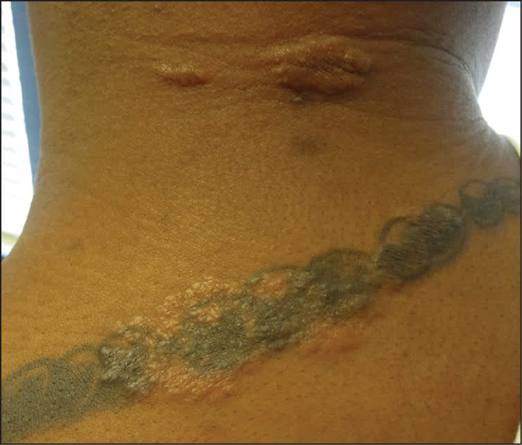
A 34-year-old man with no significant medical history presented with a rash on his arms (Figure 1) and posterior neck (Figure 2). The rash began on his left arm two years earlier after he got a tattoo. The eruption was composed of scattered papules and plaques measuring 1 to 2 cm in size. It occasionally itched, but was usually asymptomatic.
Figure 1.
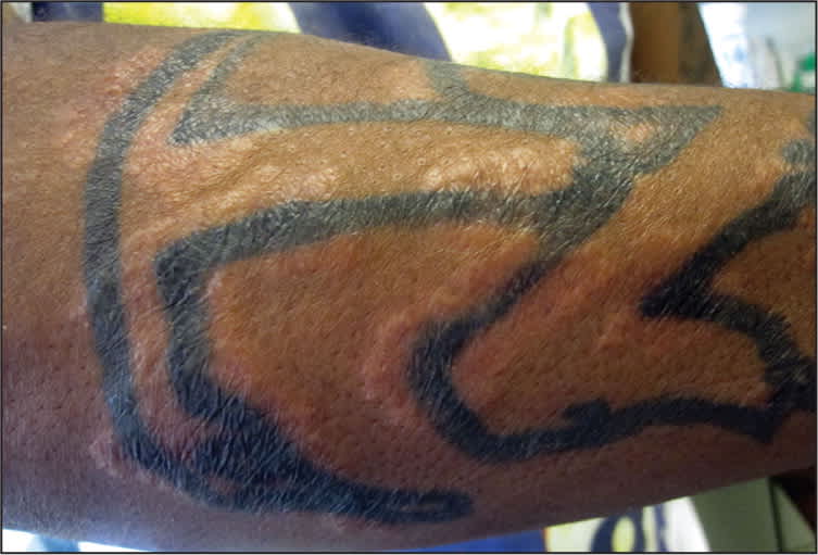
Figure 2.
Question
Based on the patient's history and physical examination, which one of the following is the most likely diagnosis?
A. Contact dermatitis.
B. Cutaneous sarcoidosis.
C. Discoid lupus erythematosus.
D. Tattoo granuloma.
Discussion
The answer is B: cutaneous sarcoidosis. Sarcoidosis is a systemic granulomatous disease that may involve the skin alone (25 percent of cases1) or other tissue or internal organs. Flesh-colored papules are the most common presentation, but many other forms exist, including lupus pernio and annular, hypopigmented, ulcerative, subcutaneous, and ichthyosiform sarcoidosis. On histologic examination, sarcoidosis often displays the classic “naked granuloma” within the dermis.
The development of numerous sarcoidal lesions in tattooed areas is a classic example of an isomorphic response, or Koebner phenomenon (i.e., the appearance of identical lesions at sites of previous trauma).1 The Koebner phenomenon may occur in a variety of other cutaneous diseases, including psoriasis, vitiligo, and lichen planus.2 Studies have suggested that increases in proinflammatory cytokines, such as tumor necrosis factor α and interleukin-1, after traumatic insult play a role in the Koebner phenomenon.3
First-line therapy for cutaneous sarcoidosis is mid-to high-potency topical corticosteroids. Additional treatment modalities include intralesional corticosteroid injection and hydroxychloroquine. Systemic sarcoidosis commonly requires long-term therapy with hydroxychloroquine and oral steroids.
Contact dermatitis manifests as exudative, extremely pruritic papules or plaques. The presence of linear or geometric-appearing eruptions suggests an external exposure or insult. For example, allergic contact dermatitis from poison ivy often exhibits linearly distributed vesicles that correspond to the sites of contact with the plant leaves. These reactions demonstrate epidermal spongiosis on biopsy rather than the granulomatous reaction seen in sarcoidosis.4
Discoid lupus erythematosus is a form of cutaneous lupus erythematosus that tends to affect the face, scalp, and ears. The rash may appear as pink to red papules and plaques. Chronic lesions become hyperpigmented or hypopigmented and atrophic, and lead to a scarring alopecia if they involve areas of hair growth. Discoid lupus erythematosus may be clinically indistinguishable from sarcoidosis; however, on histologic examination, discoid lupus erythematosus demonstrates atrophy with an interface dermatitis involving the basal layer of the epidermis.5
Tattoo granulomas are a granulomatous hypersensitivity reaction to the pigment used in tattooing. These lesions are often clinically indistinguishable from sarcoidosis, but they occur only in areas of tattooing. Histologic evaluation reveals granulomas with pigment-laden macrophages.1
Summary Table
| Condition | Characteristics |
|---|---|
| Contact dermatitis | Exudative, extremely pruritic papules or plaques in a linear or geometric-appearing distribution; histologic examination shows epidermal spongiosis |
| Cutaneous sarcoidosis | Lesions typically appear as flesh-colored papules; may demonstrate koebnerization; histologic examination demonstrates “naked granulomas” |
| Discoid lupus erythematosus | Pink to red papules and plaques that become hyperpigmented or hypopigmented and atrophy; tends to affect the face, scalp, and ears; histologic examination demonstrates atrophy with an interface dermatitis involving the basal layer of the epidermis |
| Tattoo granuloma | Lesions occurring only at the site of a tattoo; histologic examination demonstrates granulomas with pigment-laden macrophages |
