A 27-year-old otherwise healthy veterinary student presented with a slow-growing, painless, red mass on his finger that first appeared three weeks earlier. Weeping occurred without provocation, and it bled lightly with minimal trauma. It did not respond to an over-the-counter antibiotic ointment. The patient reported a minor skin break in the same area and that he had intubated a goat without wearing gloves about one month before the development of the lesion. He had no other local symptoms or history of an insect bite, fever, or malaise.
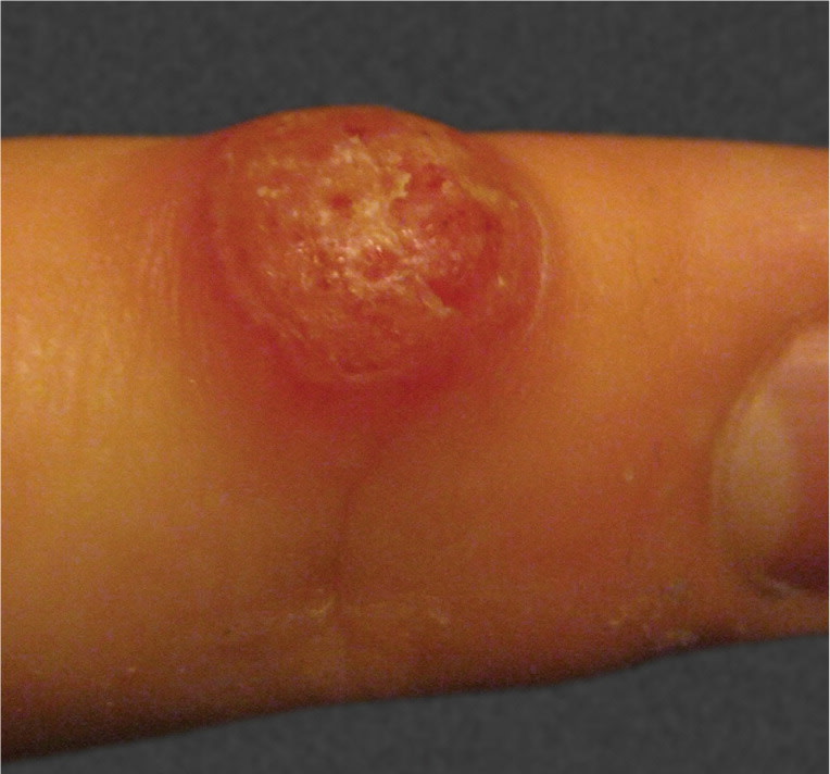
Physical examination revealed a 1-cm, well-circumscribed, erythematous nodule located on the dorsolateral aspect of the distal third finger (see accompanying figure). The lesion was firm and nontender to palpation. There was scant overlying exudate, but no surrounding erythema, edema, regional lymphadenopathy, or lymphangitis.
Question
Based on the patient’s history and physical examination findings, which one of the following is the most likely diagnosis?
A. Anthrax.
B. Milker’s nodule.
C. Orf.
D. Pyogenic granuloma.
E. Tularemia.
Discussion
The answer is C: orf. Orf, or contagious ecthyma, is a cutaneous zoonosis caused by a DNA virus of the Parapoxvirus genus that is endemic in sheep and goats. Transmission to humans usually occurs through direct contact with an infected animal (facilitated by a break in the skin), and less commonly by contacting contaminated meat, hides, or fomites.1 Orf is a common occupational disease, typically affecting the dorsum of the hands and fingers of farmers, butchers, and veterinarians.
The diagnosis of orf is clinical, based on a classic history and physical examination. Following a three- to seven-day incubation period, the typically painless lesion progresses through six well-described, one-week stages.1 This may be accompanied by low-grade fever and regional lymphadenopathy. The first (maculopapular) stage begins with a red macule at the inoculation site that progresses into a papule, which changes in appearance during subsequent stages. The second (targetoid) stage is characterized by an inner white ring with an outer red halo. The third (active) stage involves expansion and weeping, which is depicted in the accompanying figure. In the fourth (regenerative) stage, the papule is dry with a thin crust of black dots. In the fifth (papillomatous) stage, the surface becomes verrucous. The sixth (regressive) stage ends with thick crusting and spontaneous resolution, usually without scarring.
The treatment of orf is generally supportive (e.g., nonsteroidal anti-inflammatory drugs, moist dressings, immobilization) because the lesions resolve spontaneously after about six weeks. Cryotherapy may hasten recovery but typically is not indicated. Case reports show some improvement with topical antivirals, although this is not the current standard of care.1–3 Secondary bacterial infections should be treated if present.
Anthrax is a rare zoonosis caused by Bacillus anthracis, a gram-positive bacillus found in many wild and domesticated hoofed animals. Cutaneous disease is the most common and least serious form of anthrax. Transmission occurs by subcutaneous inoculation with spores originating on infected animals, their hides, or surrounding soil. Following a seven-day incubation period, a rapidly enlarging, painless, red papule emerges and progresses into a necrotic ulcer with black eschar. Extensive local edema, regional lymphadenopathy, and lymphangitis usually are present.4 Mortality drops from 20 percent to less than 1 percent with treatment.5
Milker’s nodule is a cutaneous zoonosis caused by a Parapoxvirus species endemic to cattle. Other than a different exposure history, the presentation, course, and treatment of milker’s nodule are essentially identical to orf. Definitive differentiation is unnecessary, but can be accomplished through viral culture, polymerase chain reaction, or histopathologic examination.
Pyogenic granuloma is a noninfectious capillary proliferation secondary to a minor skin insult. It presents as a rapidly enlarging, soft, red papule that bleeds easily and occasionally profusely.4 This is the most common misdiagnosis in a patient with orf.
Tularemia is a relatively rare cutaneous zoonosis caused by Francisella tularensis, a highly virulent, gram-negative coccobacillus found in rabbits and rodents. Transmission usually occurs through an arthropod vector rather than direct animal contact. A ulceroglandular presentation is most common and is characterized by a single rapidly enlarging, painful, red papule that develops a central necrotic ulcer and black eschar. Fever and regional lymphadenopathy usually are present.4,6
Summary Table
| Condition | Transmission | Characteristics |
|---|---|---|
| Anthrax | Bacillus anthracis infection from direct contact with spores originating on hoofed animals, their hides, or surrounding soil | Rapidly enlarging, painless, red papule that ulcerates and forms a black eschar; extensive local edema, regional lymphadenopathy, and lymphadenitis usually are present |
| Milker’s nodule | Parapoxvirus infection from direct contact with cattle | Self-limited, painless, red papule that evolves through six stages; presentation and course essentially identical to orf |
| Orf | Parapoxvirus infection from direct contact with sheep or goats | Self-limited, painless, red papule that evolves through six stages |
| Pyogenic granuloma | Noninfectious capillary proliferation secondary to minor skin insult | Rapidly enlarging, soft, red papule that bleeds easily |
| Tularemia | Francisella tularensis infection from rabbits and rodents via arthropod vectors | Single rapidly enlarging, painful, red papule; ulcerates and forms a black eschar; fever and regional lymphadenopathy usually present |
