A 28-year-old man presented with a rash that originated on his left arm one week earlier. The rash was very pruritic, especially in the evenings. Although the patient applied over-the-counter steroid cream, the itching continued, and the rash spread to his right arm, chest, and lower extremities. He had no personal or family history of skin disorders, including eczema or psoriasis. The patient had not been exposed to any new soaps, detergents, medications, deodorants, other personal hygiene items, plants, animals, or chemicals.
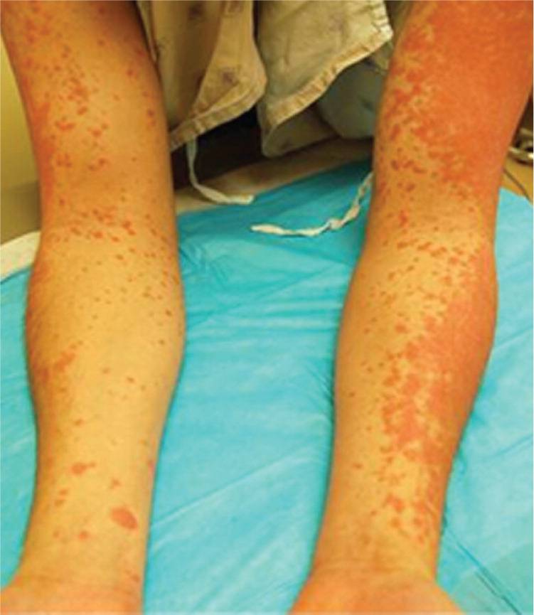
The physical examination revealed a red, sharply defined rash with scaling papules that had coalesced to form large blanching plaques over the dorsal forearms, anterior shins, and lateral ankles. The ventral forearms, chest, and stomach had numerous erythematous papules ranging from 0.2 mm to 1.0 cm in size (Figures 1 and 2). The palms and soles were spared.
Figure 1.
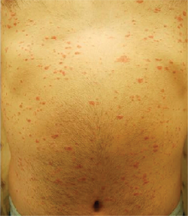
Figure 2.
Question
Based on the patient's history and physical examination, which one of the following is the most likely diagnosis?
A. Eczema.
B. Guttate psoriasis.
C. Pityriasis rosea.
D. Tinea corporis.
E. Tinea versicolor.
Discussion
The answer is B: guttate psoriasis. Guttate psoriasis can develop at any age, although it is most common in children, and occurs following group A streptococcal pharyngitis. The plaques can be up to 1 cm in size and appear soon after the sore throat begins. They are most common on the trunk and extremities, but spare the palms and soles. A history of a recent sore throat or gastrointestinal illness, and positive results on a rapid strep test or throat culture, can be helpful in making the diagnosis. This patient's rapid strep test and throat culture were positive for group A streptococcus.1
The treatment for guttate psoriasis includes treating the streptococcal infection with penicillin, and treating the skin lesions with topical triamcinolone 0.1% cream twice daily and ultraviolet B therapy.1
The scaling that occurs with eczema is more ill-defined and may have a “craquele” (cracked) appearance, with excoriation and lichenification. The lesions in acute eczematous inflammation may weep or ooze a clear fluid, and pruritus is often severe. In contrast to eczema, psoriasis typically has a red, silvery, adherent scale with sharply defined borders; therefore, pinpoint bleeding occurs when the scale is removed. Eczema plaques usually first appear on the knees, elbows, and scalp.1
Pityriasis rosea is usually asymptomatic, and most often occurs after a mild upper respiratory tract infection. The lesions may start with a single lesion on the trunk (herald patch), with numerous lesions appearing suddenly after one to two weeks. This rash is often described as scaly plaques with an inward-facing scale, called a collarette. The lesions tend to be oval, and the long axis follows relaxed skin tension lines. Therefore, on the back, the rash resembles a “Christmas tree” distribution.1
Tinea corporis is a dermatophyte infection that can appear anywhere on the body. It tends to occur more often in warm, humid climates. The lesions appear as annular, flat papules with a scaly raised border. The plaques can range from a few centimeters to several inches in size, but are always characterized by the slowly advancing border. Fungal infections can invade deep into the hair follicle and produce red papules and pustules. Microscopic examinations of a sample from the scales with a potassium hydroxide preparation show abundant hyphae.1
The lesions of tinea versicolor are small, circular, scaling papules. They are most common on the upper trunk but can extend to the bilateral arms, neck, and abdomen. The lesions are pink in fair-skinned persons, but are hypopigmented in those with tanned skin.1
Summary Table
| Condition | Characteristics |
|---|---|
| Eczema | Ill-defined, “craquele” (cracked) appearance with excoriation and lichenification; severe pruritus |
| Guttate psoriasis | Follows group A streptococcal pharyngitis, starting one to two weeks after the sore throat begins; widespread plaques spare the palms and soles |
| Pityriasis rosea | Most common after an upper respiratory tract infection; begins as a herald patch with numerous lesions appearing suddenly after one to two weeks; inward- facing scale (collarette); follows relaxed skin tension lines; “Christmas tree” distribution on the back |
| Tinea corporis | Occurs anywhere on the body; more common in warm, humid climates; annular, flat papules with a scaly, raised, slowly advancing border; potassium hydroxide preparation positive for abundant hyphae |
| Tinea versicolor | Small, circular papules; pink lesions on fair skin, hypopigmented lesions on tanned skin |
