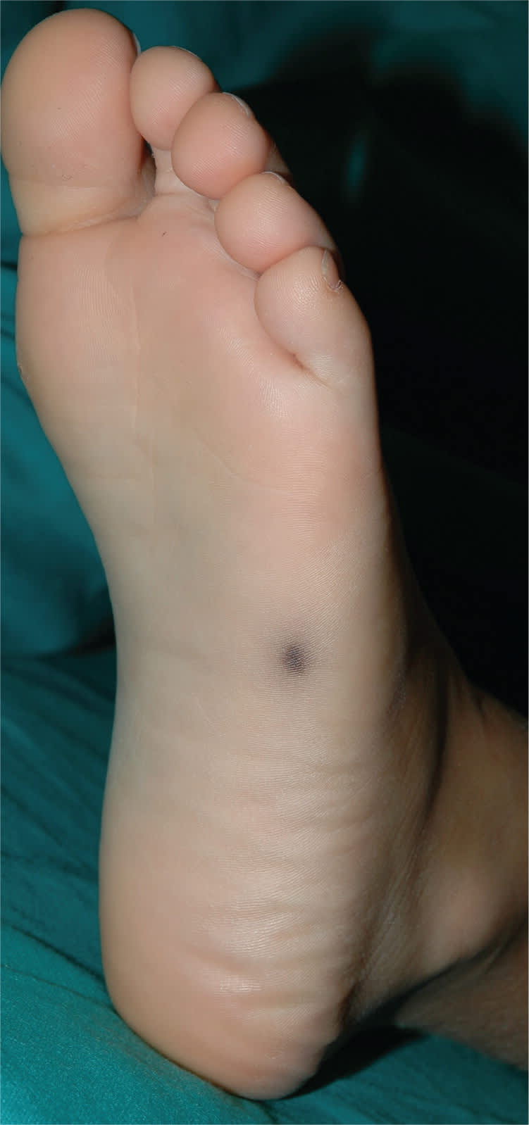
A six-year-old boy had a pigmented lesion on the sole of his foot that appeared one month earlier. He had no chronic medical problems.
[ corrected] Physical examination showed an asymptomatic brown lesion measuring 7 mm (see accompanying figure). The margins were not well defined, and a light-gray halo surrounded the lesion. A scratch test of the stratum corneum partially removed the pigmentation.
Question
Based on the patient's history and physical examination findings, which one of the following is the most likely diagnosis?
A. Acral melanoma.
B. Blue nevus.
C. Junctional melanocytic nevus.
D. Mycosis.
E. Subcorneal hematoma.
Discussion
The answer is E: subcorneal hematoma. Subcorneal hematomas are caused by traumatic rupture of the papillary capillaries, leading to extravasation of blood into the cornified layer of the sole of the foot. It can be difficult to clinically differentiate subcorneal hematoma from acral melanoma or melanocytic nevi. In children, it is especially difficult to connect the appearance of the lesion to a specific injury.
Dermoscopy can assist in the diagnosis.1,2 The presence of a red-black, homogeneous pigmentation combined with satellite globules (the light-gray halo) is a distinctive feature of subcorneal hematoma.3 The lesion is well-demarcated; round or irregularly shaped; and sometimes linear, macular, or nodular. A scratch test is another useful tool for the diagnosis of subcorneal hematoma and to exclude melanoma or melanocytic nevi. The scratch test involves using a scalpel to remove the erythrocytes located in the stratum corneum, which results in complete or partial removal of the lesion.
If the clinical history and dermoscopic features of a pigmented lesion are not diagnostic and the results of a scratch test are negative, the diagnosis should be confirmed by surgical excision and histopathologic examination. Subcorneal hematomas regress spontaneously after a few months.
A rapidly growing pigmented lesion may suggest malignant acral melanoma. An acral melanoma may initially present as a 5- to 12-mm, asymmetrical, flattened plaque with uneven edges, irregular borders, dark-brown or black color, and uneven pigmentation. In the advanced phase of vertical growth, it may look papular or nodular.
A blue nevus typically presents as a blue, blue-gray, or blue-black papule or nodule less than 1 cm. However, blue nevi usually do not change in appearance over time.
A junctional melanocytic nevus typically appears as a macula that is smaller than 1 cm. It is brown with uniform pigmentation, has a round or oval shape, and has smooth edges. The lesion usually does not regress spontaneously.
Cutaneous or subcutaneous mycosis usually leads to erythematous or desquamative plaques or nodules. They can be single or multiple, and are located at the site of infection, often on the extremities or other exposed areas. They are usually slow growing and do not regress spontaneously.4
Summary Table
| Condition | Characteristics |
|---|---|
| Acral melanoma | Rapidly growing, asymmetrical, flattened plaque; 5 to 12 mm; uneven edges, irregular borders, dark-brown or black color, and uneven pigmentation |
| Blue nevus | Blue, blue-gray, or blue-black papule or nodule; < 1 cm; appearance usually does not change |
| Junctional melanocytic nevus | Brown macule; < 1 cm; uniform pigmentation, round or oval shape, and smooth edges; generally does not regress spontaneously |
| Cutaneous or subcutaneous mycosis | Erythematous or desquamative plaques or nodules; single or multiple; usually located on extremities or other exposed area; slow growing; generally does not regress spontaneously |
| Subcorneal hematoma | Red-black, homogeneous pigmentation with light-gray halo; well-demarcated; round or irregularly shaped; sometimes linear, macular, nodular; regresses spontaneously |
