Guideline source: American College of Physicians
Evidence rating system used? Yes
Literature search described? Yes
Guideline developed by participants without relevant financial ties to industry? No
Published source: Annals of Internal Medicine, November 20, 2012
Available at: http://annals.org/article.aspx?articleid=1392193
The American College of Physicians (ACP), in collaboration with the American College of Cardiology Foundation, American Heart Association, American Association for Thoracic Surgery, Preventive Cardiovascular Nurses Association, and Society of Thoracic Surgeons, has developed a guideline that helps physicians diagnose known or suspected cases of stable ischemic heart disease (IHD). Recommendations address IHD and related issues, including initial diagnosis, cardiac stress testing, and coronary angiography.
Initial Cardiac Testing
Patients with chest pain should undergo a thorough history and physical examination to determine the probability of IHD before undergoing additional testing. The patient and physician should participate in the decision-making process regarding diagnostic and therapeutic options, with the physician explaining information about risks, benefits, and costs of care to the patient.
Patients who present with acute angina must be categorized as stable or unstable. Patients who have unstable angina should be further categorized as high, intermediate, or low risk (Table 1). In patients with an obvious noncardiac cause of chest pain, resting electrocardiography (ECG) is recommended for risk assessment.
Table 1. Short-Term Risk of Death or Nonfatal Myocardial Infarction in Patients with Unstable Angina*
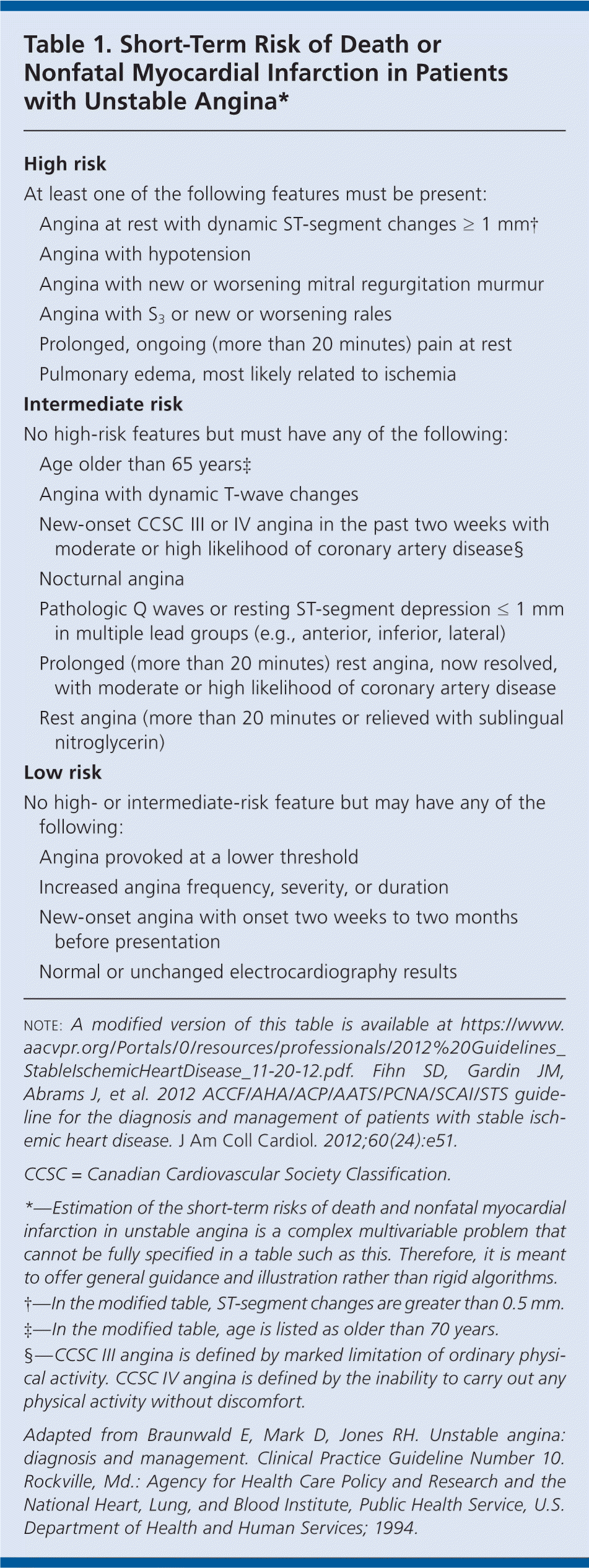
| High risk | |
| At least one of the following features must be present: | |
| Angina at rest with dynamic ST-segment changes ≥ 1 mm† | |
| Angina with hypotension | |
| Angina with new or worsening mitral regurgitation murmur | |
| Angina with S3 or new or worsening rales | |
| Prolonged, ongoing (more than 20 minutes) pain at rest | |
| Pulmonary edema, most likely related to ischemia | |
| Intermediate risk | |
| No high-risk features but must have any of the following: | |
| Age older than 65 years‡ | |
| Angina with dynamic T-wave changes | |
| New-onset CCSC III or IV angina in the past two weeks with moderate or high likelihood of coronary artery disease§ | |
| Nocturnal angina | |
| Pathologic Q waves or resting ST-segment depression ≤ 1 mm in multiple lead groups (e.g., anterior, inferior, lateral) | |
| Prolonged (more than 20 minutes) rest angina, now resolved, with moderate or high likelihood of coronary artery disease | |
| Rest angina (more than 20 minutes or relieved with sublingual nitroglycerin) | |
| Low risk | |
| No high- or intermediate-risk feature but may have any of the following: | |
| Angina provoked at a lower threshold | |
| Increased angina frequency, severity, or duration | |
| New-onset angina with onset two weeks to two months before presentation | |
| Normal or unchanged electrocardiography results | |
NOTE: A modified version of this table is available at https://www.aacvpr.org/Portals/0/resources/professionals/2012%20Guidelines_StableIschemicHeartDisease_11-20-12.pdf. Fihn SD, Gardin JM, Abrams J, et al. 2012 ACCF/AHA/ACP/AATS/PCNA/SCAI/STS guideline for the diagnosis and management of patients with stable ischemic heart disease. J Am Coll Cardiol. 2012;60(24):e51.
CCSC = Canadian Cardiovascular Society Classification.
*—Estimation of the short-term risks of death and nonfatal myocardial infarction in unstable angina is a complex multivariable problem that cannot be fully specified in a table such as this. Therefore, it is meant to offer general guidance and illustration rather than rigid algorithms.
†—In the modified table, ST-segment changes are greater than 0.5 mm.
‡—In the modified table, age is listed as older than 70 years.
§—CCSC III angina is defined by marked limitation of ordinary physical activity. CCSC IV angina is defined by the inability to carry out any physical activity without discomfort.
Adapted from Braunwald E, Mark D, Jones RH. Unstable angina: diagnosis and management. Clinical Practice Guideline Number 10. Rockville, Md.: Agency for Health Care Policy and Research and the National Heart, Lung, and Blood Institute, Public Health Service, U.S. Department of Health and Human Services; 1994.
In patients with an intermediate pretest probability of IHD who have interpretable ECG results and at least moderate physical functioning or no disabling comorbidity, standard exercise ECG is recommended for initial diagnosis. In patients with an intermediate to high pretest probability of IHD whose ECG results cannot be interpreted and who have at least moderate physical functioning or no disabling comorbidity, exercise stress testing with radionuclide myocardial perfusion imaging or echocardiography should be used.
Pharmacologic stress testing with radionuclide myocardial perfusion imaging, echocardiography, or cardiac magnetic resonance imaging should not be used for patients who have interpretable ECG results and at least moderate physical functioning or no disabling comorbidity. Exercise stress testing with nuclear myocardial perfusion imaging should not be used as an initial test in low-risk patients who have these same criteria.
Pharmacologic stress testing with radionuclide myocardial perfusion imaging or echocardiography is recommended in patients with an intermediate to high pretest probability of IHD who are incapable of at least moderate physical functioning or who have a disabling comorbidity.
Standard exercise ECG testing should not be used for patients who have ECG results that cannot be interpreted, who are incapable of at least moderate physical functioning, or who have a disabling comorbidity.
Assessing resting left ventricular systolic and diastolic function and evaluating for abnormalities of myocardium, pericardium, or heart valves using Doppler echocardiography are recommended in patients who have known or suspected IHD and previous myocardial infarction, pathologic Q waves, signs or symptoms that suggest heart failure, complex ventricular arrhythmias, or an undiagnosed heart murmur.
Cardiac computed tomography, cardiac magnetic resonance imaging, echocardiography, and radionuclide imaging should not be used for routine assessment of left ventricular function in patients who have normal ECG results, no history of myocardial infarction, no signs or symptoms that suggest heart failure, and no complex ventricular arrhythmias. Routine reassessment (less than one year) of left ventricular function using these tests is inappropriate in patients who have no change in clinical status and for whom no change in therapy is contemplated.
Cardiac Stress Testing in Known Stable IHD
Standard exercise ECG testing for risk assessment is recommended in patients with known stable IHD who are able to exercise and have ECG results that can be interpreted during exercise. If patients can exercise but have uninterpretable ECG results not caused by left bundle branch block or ventricular pacing, adding radionuclide myocardial perfusion imaging or echocardiography to standard exercise ECG testing is recommended. Pharmacologic stress testing or cardiac computed tomographic angiography should not be used to assess risk in patients with stable IHD who are able to exercise and have interpretable ECG results.
In patients with known stable IHD who are unable to exercise regardless of the ability to interpret the patient's ECG results, pharmacologic stress testing with radionuclide myocardial perfusion imaging or echocardiography is recommended. Regardless of the patient's ability to exercise, pharmacologic stress testing with radionuclide myocardial perfusion imaging or echocardiography for risk assessment in patients with stable IHD who have left bundle branch block on ECG is recommended.
In patients being considered for revascularization of known coronary stenosis of unclear physiologic significance, exercise or pharmacologic stress testing with imaging for risk assessment is recommended.
More than one stress imaging study, or a stress imaging study and cardiac computed tomography angiography at the same time, should not be used for risk assessment in patients with stable IHD.
Coronary Angiography
Patients with stable IHD who have survived sudden cardiac death or potentially life-threatening ventricular arrhythmia should undergo coronary angiography to assess cardiac risk. Patients with stable IHD who develop signs and symptoms of heart failure should be evaluated to determine if coronary angiography should be performed. Patients with stable IHD and clinical features indicative of a high likelihood of severe IHD should have coronary angiography to determine cardiac risk.
Coronary angiography should be used for risk assessment in patients with stable IHD whose clinical characteristics and results of noninvasive testing indicate a high likelihood of severe IHD, and when the benefits of coronary angiography outweigh the risks.
Coronary angiography should not be used to assess risk in patients with stable IHD who decline revascularization, or who are not candidates for revascularization based on comorbidities or individual preferences; to further assess risk in patients with stable IHD who have preserved left ventricular function and low-risk criteria on noninvasive testing; for risk assessment in patients who are at low risk based on clinical criteria and who have not undergone noninvasive risk testing; or in asymptomatic patients with no indication of ischemia on noninvasive testing.
