A 62-year-old woman presented with a four-week history of edema in both of her upper extremities, as well as changes in skin pigmentation. She had alopecia involving the frontal area of the scalp. She had dyspnea on exertion, which had worsened over several weeks.
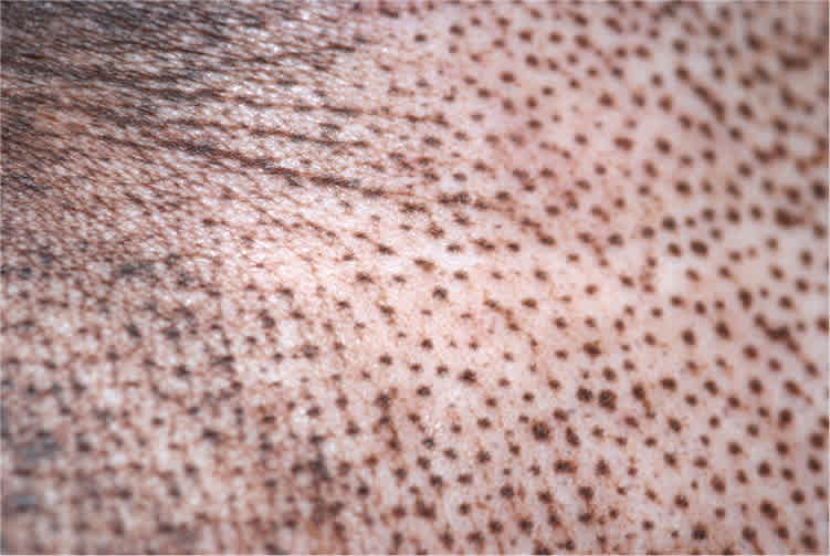
Physical examination showed firm skin with nonpitting edema in the forearms, hands, and fingers. She also had pigmentation changes on her face, neck, and upper back (Figures 1 through 3).
Figure 1.
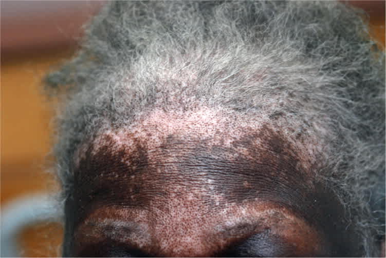
Figure 2.
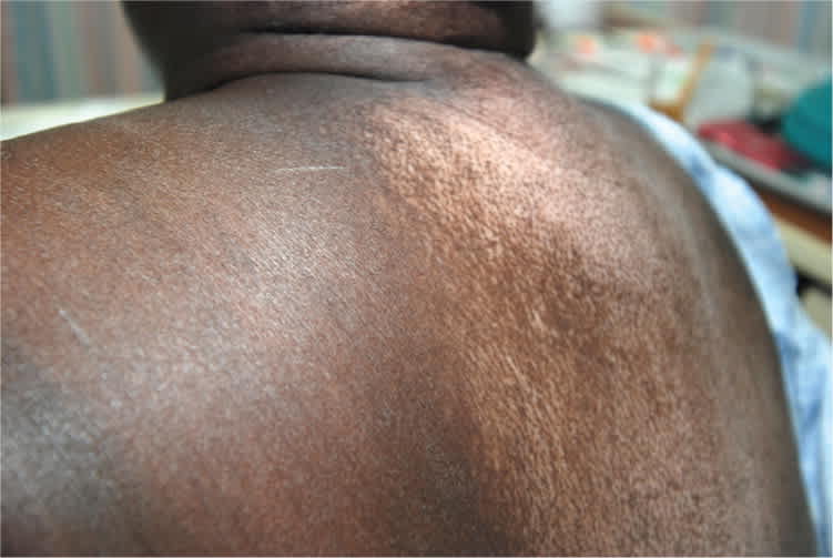
Figure 3.
Laboratory results included the following: white blood cell count, 10,000 per mm3 (10.0 × 10⁹ per L); hemoglobin, 8.5 g per dL (85 g per L); erythrocyte sedimentation rate, 115 mm per hour (normal, 0 to 30 mm per hour); sodium, 137 mEq per L (137 mmol per L); potassium, 4.4 mEq per L (4.4 mmol per L); blood urea nitrogen, 23 mg per dL (8.2 mmol per L); creatinine, 1.4 mg per dL (124 μmol per L); urinalysis, normal; antinuclear antibody, positive (1:640) with a nucleolar pattern; and rheumatoid factor, 24.6 IU per mL (normal, 0 to 13.9 IU per mL). Chest radiography showed cardiomegaly with evidence of vascular congestion.
Question
Based on the patient's history, physical examination, and laboratory findings, which one of the following is the most likely diagnosis?
A. Dermatomyositis.
B. Rheumatoid arthritis.
C. Scleroderma.
D. Systemic lupus erythematosus.
E. Vitiligo with repigmentation.
Discussion
The correct answer is C: scleroderma. Nonpitting edema may appear months or years before the characteristic changes of scleroderma develop.1 These changes may include tightly bound skin, with ulcer formation, tightness of the facial skin with fine perioral wrinkling, and an inability to fully open the mouth. The skin changes tend to affect fingers and hands initially, with gradual proximal spread to the forearms.1 The lower extremities tend to be relatively spared.1
“Salt-and-pepper” pigmentation changes are characteristic of scleroderma with perifollicular sparing of pigment. This pattern is most common on the scalp, upper back, and chest.2 Dermal sclerosis may obliterate hair follicles, resulting in hair loss. Pulmonary involvement may include interstitial lung disease and pulmonary artery hypertension.2 Nucleolar pattern antinuclear antibodies are associated with antiribonucleoprotein, which is characteristic of scleroderma and rare in other immune disorders.3
Dermatomyositis often presents as proximal muscle weakness and a heliotrope rash, commonly involving the eyelids.4
Rheumatoid arthritis is typically associated with symmetrical joint involvement, morning stiffness, synovial thickening, and a family history of the disease.2
Systemic lupus erythematosus can manifest in a variety of ways. Dermatologic manifestations include nonpruritic, erythematous to violaceous plaques, often on sun-exposed areas. Telangiectasias, alopecia, urticaria, and Raynaud phenomenon are also common.4
Vitiligo with repigmentation can appear similarly to this patient's presentation, with areas of repigmentation beginning in the perifollicular area. Although vitiligo has been associated with several other conditions (e.g., hypothyroidism, Graves disease, Addison disease, pernicious anemia, diabetes mellitus), most patients with vitiligo have no other associated findings.4
Summary Table
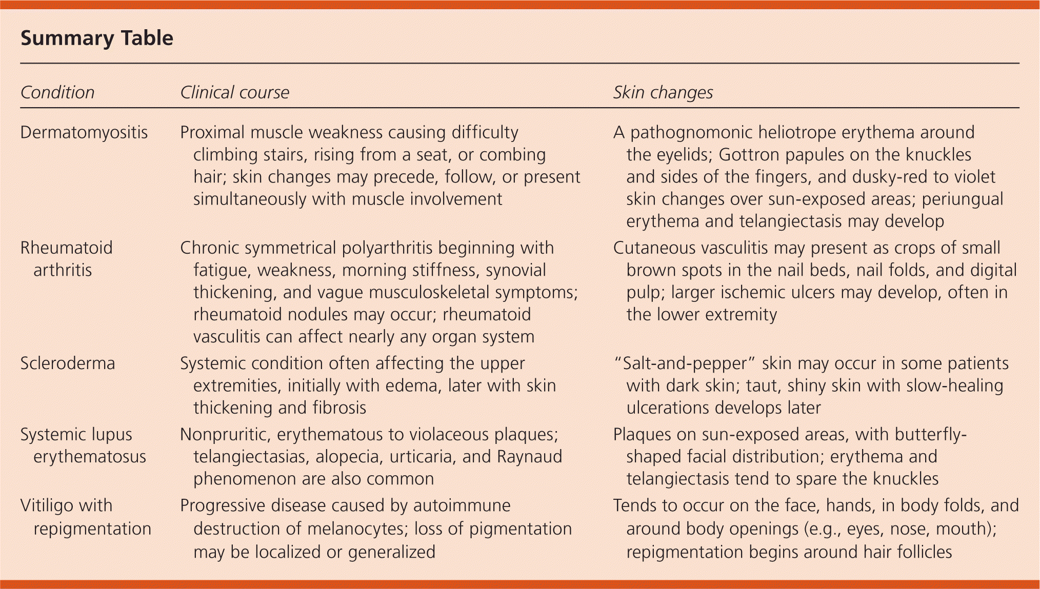
| Condition | Clinical course | Skin changes |
|---|---|---|
| Dermatomyositis | Proximal muscle weakness causing difficulty climbing stairs, rising from a seat, or combing hair; skin changes may precede, follow, or present simultaneously with muscle involvement | A pathognomonic heliotrope erythema around the eyelids; Gottron papules on the knuckles and sides of the fingers, and dusky-red to violet skin changes over sun-exposed areas; periungual erythema and telangiectasis may develop |
| Rheumatoid arthritis | Chronic symmetrical polyarthritis beginning with fatigue, weakness, morning stiffness, synovial thickening, and vague musculoskeletal symptoms; rheumatoid nodules may occur; rheumatoid vasculitis can affect nearly any organ system | Cutaneous vasculitis may present as crops of small brown spots in the nail beds, nail folds, and digital pulp; larger ischemic ulcers may develop, often in the lower extremity |
| Scleroderma | Systemic condition often affecting the upper extremities, initially with edema, later with skin thickening and fibrosis | “Salt-and-pepper” skin may occur in some patients with dark skin; taut, shiny skin with slow-healing ulcerations develops later |
| Systemic lupus erythematosus | Nonpruritic, erythematous to violaceous plaques; telangiectasias, alopecia, urticaria, and Raynaud phenomenon are also common | Plaques on sun-exposed areas, with butterfly-shaped facial distribution; erythema and telangiectasis tend to spare the knuckles |
| Vitiligo with repigmentation | Progressive disease caused by autoimmune destruction of melanocytes; loss of pigmentation may be localized or generalized | Tends to occur on the face, hands, in body folds, and around body openings (e.g., eyes, nose, mouth); repigmentation begins around hair follicles |
