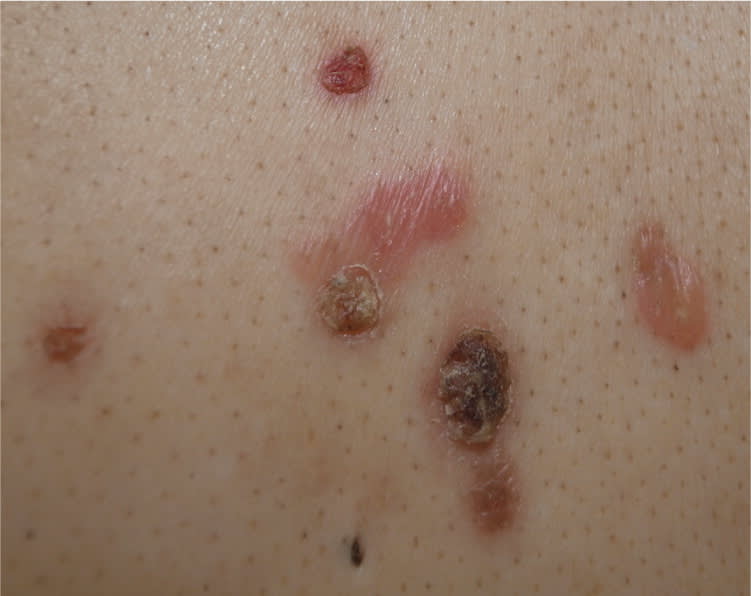
A 62-year-old man with no significant medical history presented with multiple bullae on his trunk that had been present for one week. The rash was painful but not pruritic. He did not have a fever or sore throat, and had not used any new medications.
Physical examination revealed multiple vesicles, flaccid bullae, and erosions with crust formation on the chest, abdomen, and back (Figures 1 and 2). Mucosal erosions and the Nikolsky sign were also present. There was no lymphadenopathy. Biopsy of a blister showed suprabasal acantholysis with intraepidermal blister formation and follicular involvement. Direct immunofluorescence revealed intercellular deposits of immunoglobulin G and C3 within the epidermis.
Figure 1.
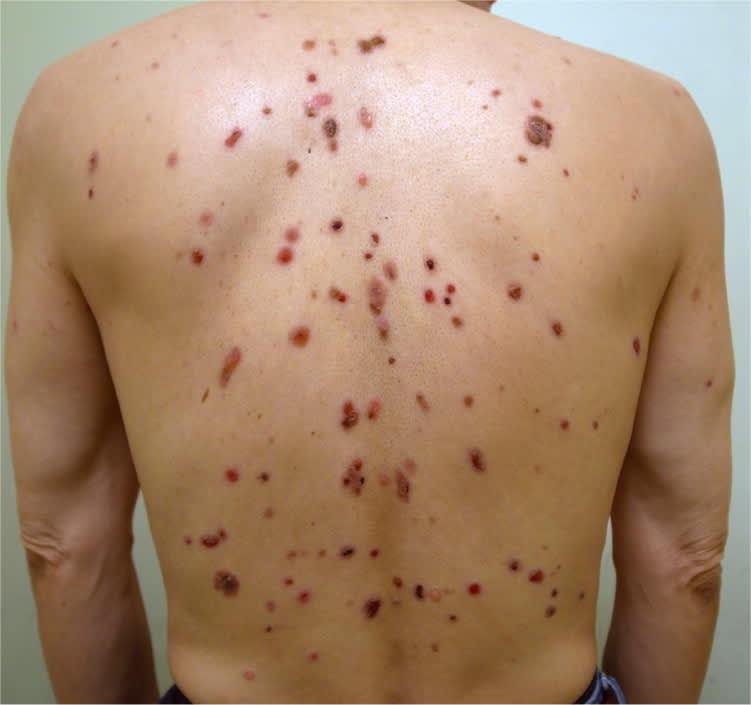
Figure 2.
Question
Based on the patient's history and physical examination findings, which one of the following is the most likely diagnosis?
A. Bullous impetigo.
B. Bullous pemphigoid.
C. Dermatitis herpetiformis.
D. Pemphigus vulgaris.
Discussion
The answer is D: pemphigus vulgaris. Pemphigus vulgaris is an autoimmune blistering disease of the skin and mucous membranes. It presents as flaccid, easily ruptured blisters arising on normal skin, with subsequent erosions and crust formation. It often appears on the face, scalp, trunk, intertriginous areas, and mucosal areas. The lesions are usually painful but rarely pruritic. The mean age of onset is 40 to 60 years. The Nikolsky sign is typically present.
Pemphigus vulgaris is caused by the loss of the normal cell-to-cell adhesion in the epidermis because of autoantibodies against desmogleins. The histologic findings are suprabasal acantholysis with intraepidermal blister formation and frequent follicular involvement. Direct immunofluorescence reveals intercellular deposits of immunoglobulin G and/or C3 within the epidermis. Detection of circulating antibodies against desmogleins by indirect immunof luorescence or enzyme-linked immunosorbent assay may help to make a diagnosis.
Myasthenia gravis and thymoma are associated with pemphigus vulgaris. Without treatment, pemphigus vulgaris leads to death; however, the mortality rate is markedly reduced with the use of systemic steroids and immunosuppressive agents.1
Bullous impetigo is a superficial cutaneous infection caused by Staphylococcus aureus. It most commonly affects neonates and infants, but also occurs in older children and adults. The thin-walled, flaccid bullae usually arise on grossly normal skin in perioral and perinasal areas, and in body folds. The bullae contain clear yellow fluid that subsequently turns turbid and dark yellow. Brown to yellowish crusts may form after the bullae rupture. Patients may have fever and malaise. Histologically, patients have subcorneal blisters filled with neutrophils, gram-positive cocci, and occasional acantholytic cells.1,2
Bullous pemphigoid is an autoimmune disease characterized by tense blisters filled with serous or hemorrhagic fluid. The blisters usually appear on the extremities and trunk, although widespread eruption is possible. Mucosal lesions occur in approximately 10% to 35% of patients. Urticarial papules and plaques may predominate in the early stage of the disease. There is usually marked pruritus. The typical age of onset is 60 to 80 years. Histologically, patients have a subepidermal blister with dermal infiltrates of lymphocytes and eosinophils. Direct immunofluorescence shows linear deposits of immunoglobulin G and C3 along the dermoepidermal junction.1
Dermatitis herpetiformis is an autoimmune disorder associated with gluten sensitivity. It presents as intensely pruritic papulovesicles symmetrically located on the extensor surfaces and buttocks. The characteristic microscopic feature is microabscesses containing neutrophils in the dermal papillary tips. Direct immunofluorescence showing immunoglobulin A deposits at the dermoepidermal junction is pathognomonic.3
Summary Table
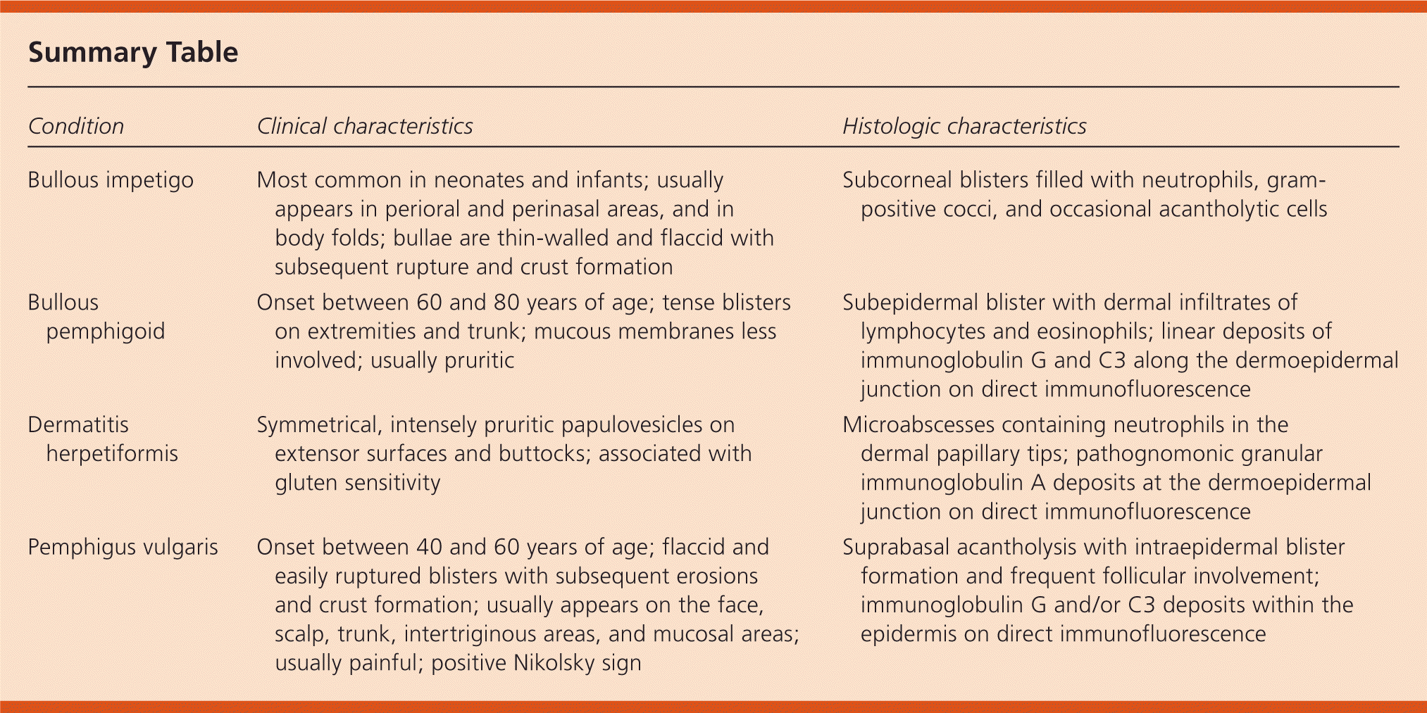
| Condition | Clinical characteristics | Histologic characteristics |
|---|---|---|
| Bullous impetigo | Most common in neonates and infants; usually appears in perioral and perinasal areas, and in body folds; bullae are thin-walled and flaccid with subsequent rupture and crust formation | Subcorneal blisters filled with neutrophils, gram-positive cocci, and occasional acantholytic cells |
| Bullous pemphigoid | Onset between 60 and 80 years of age; tense blisters on extremities and trunk; mucous membranes less involved; usually pruritic | Subepidermal blister with dermal infiltrates of lymphocytes and eosinophils; linear deposits of immunoglobulin G and C3 along the dermoepidermal junction on direct immunofluorescence |
| Dermatitis herpetiformis | Symmetrical, intensely pruritic papulovesicles on extensor surfaces and buttocks; associated with gluten sensitivity | Microabscesses containing neutrophils in the dermal papillary tips; pathognomonic granular immunoglobulin A deposits at the dermoepidermal junction on direct immunofluorescence |
| Pemphigus vulgaris | Onset between 40 and 60 years of age; flaccid and easily ruptured blisters with subsequent erosions and crust formation; usually appears on the face, scalp, trunk, intertriginous areas, and mucosal areas; usually painful; positive Nikolsky sign | Suprabasal acantholysis with intraepidermal blister formation and frequent follicular involvement; immunoglobulin G and/or C3 deposits within the epidermis on direct immunofluorescence |
