A 13-year-old girl presented with a painless bump on her left medial knee that had been present for about two weeks. She did not report any trauma or injury, and the bump was not interfering with her daily activities. Her personal and family histories were unremarkable.
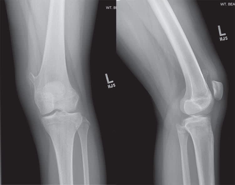
Physical examination revealed no ecchymosis or knee joint effusion. On palpation, there was a nontender bony prominence in the medial distal left femur. There was no palpable inguinal lymphadenopathy, and her knee range of motion was normal. Plain radiography was performed (Figure 1).
Figure 1.
Question
Based on the patient's history, physical examination, and radiography findings, which one of the following is the most likely diagnosis?
A. Enchondroma.
B. Fibrous dysplasia.
C. Nonossifying fibroma.
D. Osteochondroma.
E. Osteoid osteoma.
Discussion
The answer is D: osteochondroma. Osteochondromas are the most common benign bone tumors. They usually affect adolescents and children, but occur in up to 3% of the general population and account for up to 30% of all bone tumors.1 Osteochondromas are generally asymptomatic and found incidentally on imaging, but they occasionally present as painless masses.1,2 They rarely present as painful lesions due to irritation of the surrounding soft tissue.1,3 Solitary osteochondromas generally are found in the metaphysis of long bones, including the femur or tibia, but can be found in any bone.1–3 Osteochondromas usually communicate with the intramedullary bone canal.
On radiography, osteochondromas appear as bony outgrowths in continuity of the cortex and should not have cystic lesions, surrounding edema, or periostitis.1,3 Magnetic resonance imaging may be indicated for further evaluation in symptomatic cases.2 Osteochondromas usually have a cap of hyaline cartilage that is seen best on magnetic resonance imaging.2 They rarely transform into malignant lesions.1–3 Treatment is not indicated unless there is inflammation of the overlying soft tissue, risk of fracture because of the size of the osteochondroma, or mechanical joint restriction.1,3
Enchondromas are common benign bone tumors affecting all age groups and are usually found incidentally on radiography.3 They are typically asymptomatic but may be painful because of irritation of the overlying soft tissue.2,3 Enchondromas are intramedullary lesions, typically more centrally than eccentrically located, and are usually found in the metaphysis or diaphysis of long bones.2,3 On radiography, they appear benign with irregular calcification (“popcorn” or “stippled” lesion).3
Fibrous dysplasia is an uncommon benign developmental anomaly of bone formation with replacement of normal bone by a matrix of fibrous tissue and small woven spicules of bone.3,4 Fibrous dysplasias are usually localized to the metaphysis of the femur and tibia, are asymptomatic, and occur before 30 years of age.3,4 There is an increased risk of a pathologic fracture in the long bones because of bowing.3,4 Radiography shows a centrally located lesion with a ground-glass appearance of the matrix, with sharp and well-defined sclerotic margin.4
Nonossifying fibromas are common benign bone lesions found incidentally on radiography in patients younger than 30 years.5 They are most often located in the long bones, predominantly the femur and tibia.3,5 They are cortically based eccentric lesions found in the medullary cavity of long bones without periosteal reaction, irregular borders, or surrounding edema.2,3,5
Osteoid osteomas often present with pain at night overlying a hard mass, usually in male adolescents.3,5,6 Radiography shows a well-circumscribed, 1- to 2-cm lesion with a sclerotic border surrounding an osteoblastic nidus.3,6 They are usually located in the distal femur or proximal tibia.3,5,6
Summary Table
| Condition | Characteristics |
|---|---|
| Enchondroma | Benign appearance with irregular calcification (“popcorn” or “stippled” lesion); most common in the metaphysis or diaphysis of the hand, distal femur, proximal humerus, and tibia; may be painful because of irritation of the overlying soft tissue |
| Fibrous dysplasia | Ground-glass appearance of the matrix with sharp and well-defined sclerotic margin; most common in the metaphysis of the femur and tibia; bowing deformity and pathologic fracture may occur |
| Nonossifying fibroma | Geographic appearing, cortically based eccentric lesions found in the medullary cavity of long bones; usually asymptomatic and can occur before 30 years of age; more common in children and adolescents |
| Osteochondroma | Bony outgrowths that usually communicate with the intramedullary bone canal; usually occur in the metaphysis of long bones of children and adolescents |
| Osteoid osteoma | Well-circumscribed, 1- to 2-cm lesion with osteoblastic nidus and a surrounding sclerotic border; most common in male adolescents; usually located in the distal femur or proximal tibia; often causes pain at night overlying a hard mass |
