
Am Fam Physician. 2015;92(10):925-926
Author disclosure: No relevant financial affiliations.
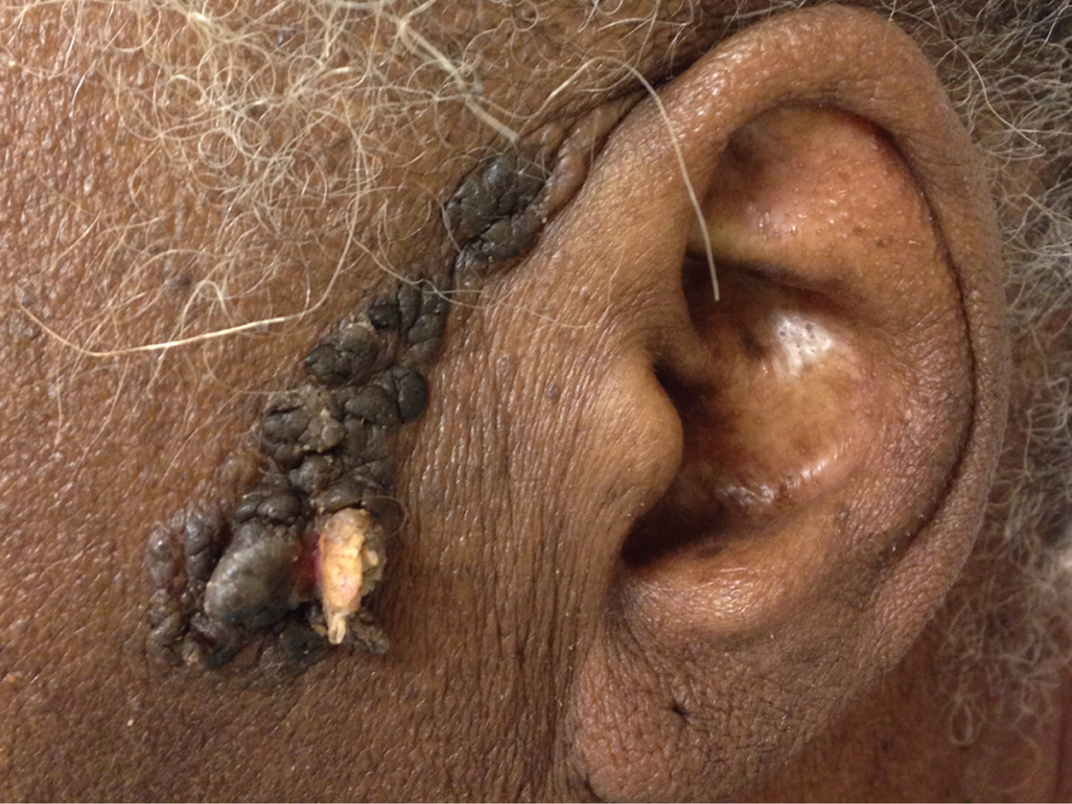
A 70-year-old black woman presented with an enlarging lesion on the left side of her face. The lesion had been present as long as the patient could remember, likely since birth. It had slowly enlarged as she aged. Recently the lesion had begun to change in appearance, and the patient developed new associated pruritus, pain, and bleeding.
The physical examination revealed a 6-cm × 1-cm linear verrucous lesion extending from the anterior helix of the left ear down to the level of the ear lobe (Figure 1). The lesion was hyperpigmented and raised, and consisted of papules and nodules. There was an area of excoriation near the distal end of the lesion where one of the nodules had torn away from the base.
Question
Discussion
The answer is C: sebaceous nevus. These lesions generally are present at birth or develop in early childhood. The most common sites of growth are the scalp and forehead. The lesions evolve along with the normal development of sebaceous glands, with changes most notable during puberty. In early childhood, the lesions are plaque-like and waxy. With pubertal changes, the lesions become larger and more verrucous.1
Secondary benign and malignant neoplasms can develop in the primary nevus, typically in middle-aged adults, although this is rare.1 An estimated 8% of sebaceous nevi develop malignant tumors,2 most commonly basal cell carcinoma. Other malignant transformations that may develop are sebaceous carcinoma, squamous cell carcinoma, and keratoacanthoma.2
Keratoacanthomas are papules with a horny keratin plug. They most commonly develop in middle or older age, and typically occur on sun-exposed areas in fair-skinned persons. They are characterized by rapid growth and involution with resolution within nine months of appearance.4
Squamous cell carcinoma most often occurs on sun-exposed areas in older, fair-skinned individuals. In dark-skinned persons, it is more likely on non–sun-exposed areas or at sites of chronic scarring. The lesions typically appear as scaly patches or plaques.6
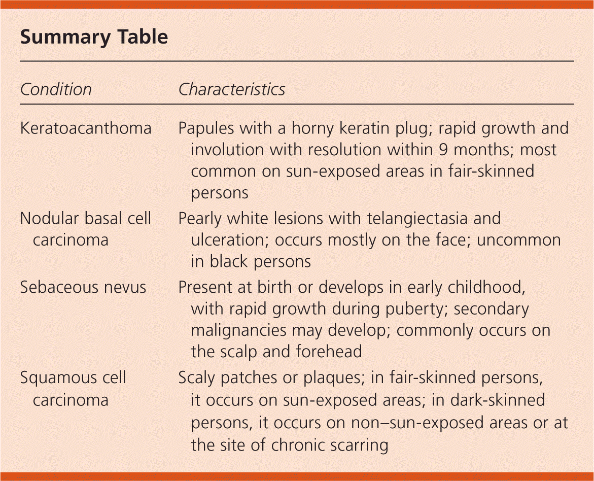
| Condition | Characteristics |
|---|---|
| Keratoacanthoma | Papules with a horny keratin plug; rapid growth and involution with resolution within 9 months; most common on sun-exposed areas in fair-skinned persons |
| Nodular basal cell carcinoma | Pearly white lesions with telangiectasia and ulceration; occurs mostly on the face; uncommon in black persons |
| Sebaceous nevus | Present at birth or develops in early childhood, with rapid growth during puberty; secondary malignancies may develop; commonly occurs on the scalp and forehead |
| Squamous cell carcinoma | Scaly patches or plaques; in fair-skinned persons, it occurs on sun-exposed areas; in dark-skinned persons, it occurs on non–sun-exposed areas or at the site of chronic scarring |
