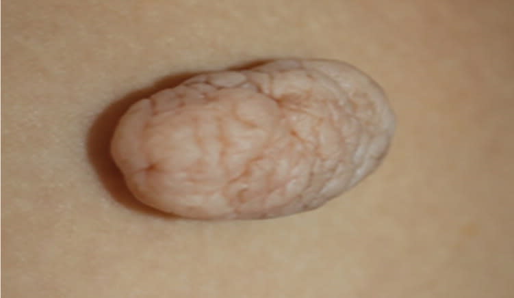
An otherwise healthy 45-year-old woman presented with a lesion on her right thigh. It had gradually increased in size since she first noticed it five years earlier. The patient had no history of trauma or surgery to the area. The lesion was asymptomatic and solitary. The physical examination revealed a well-defined, dome-shaped, corrugated, skin-colored, pedunculated mass on the right medial thigh (Figure 1, Figure 2, and Figure 3). The lesion was 3 × 4 cm in size, soft, nontender, and not reducible. The remainder of the examination was unremarkable.
Figure 1.
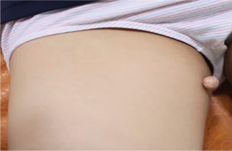
Figure 2.
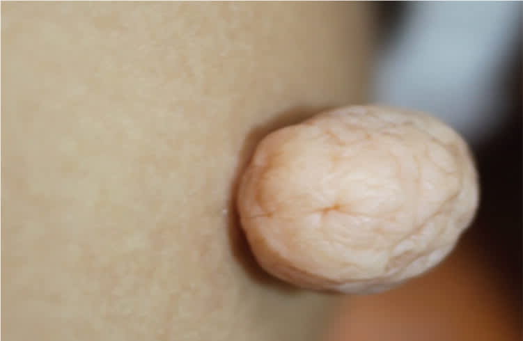
Figure 3.
Question
Based on the patient's history and physical examination findings, which one of the following is the most likely diagnosis?
A. Acrochordon.
B. Lipoma.
C. Neurofibroma.
D. Nevus lipomatosus cutaneus superficialis.
Discussion
The answer is D: nevus lipomatosus cutaneus superficialis, which is an uncommon form of benign connective tissue nevus. It is characterized by ectopic fatty tissue in the dermis. The lesions can be solitary or, more commonly, multiple. The multiple type is typically seen at birth or presents during the first three decades of life. It presents as flesh-colored or yellowish, soft papules or plaques in the pelvic girdle, most commonly the buttocks, sacrococcygeal areas, and upper posterior thighs. The solitary type, or pedunculated lipofibroma, presents as a dome-shaped, slow-growing, pedunculated mass and usually occurs after 30 years of age. The solitary form can occur on the buttocks, thighs, and nonpelvic areas, such as the axillae, arms, knees, ears, and scalp.1,2
The pathogenesis of nevus lipomatosus cutaneus superficialis is unclear. It has been hypothesized that the deposition of adipose tissue in the dermis is secondary to degenerative changes in the dermal collagen and elastic tissue, or to the displacement of subcutaneous adipose tissue into the dermis.2 Systemic abnormalities and malignant change have not been reported. Treatment is cosmetic, with surgical excision as the preferred option. Cryotherapy is an alternative treatment.1
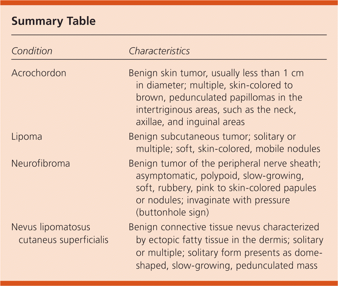
Acrochordons are also known as skin tags, soft fibromas, or fibroepithelial polyps. They are benign skin tumors that are usually less than 1 cm in diameter. They typically present as multiple, skin-colored to brown, pedunculated papillomas in the intertriginous areas, such as the neck, axillae, and inguinal areas.2 Acrochordons often increase in number when the patient is gaining weight or during pregnancy and may be related to the growth hormone–like activity of insulin. They may also occur in patients with diabetes mellitus.
Lipomas are solitary or multiple, benign, subcutaneous tumors composed of fat cells. They most often occur on the trunk, abdomen, or neck, followed by proximal extremities, but can occur anywhere. Typically, they present as soft, skin-colored, mobile nodules. Because the lesions are subcutaneous, they are not pedunculated.3
Neurofibromas are benign tumors of the peripheral nerve sheath and may present as multiple or solitary lesions. Multiple neurofibromas occur with neurofibromatosis or von Recklinghausen disease. Neurofibromas are asymptomatic, polypoid, slow-growing, soft, rubbery, pink to skin-colored papules or nodules that may vary in size.4 Larger lesions may become pedunculated over time. With pressure, the lesions invaginate (buttonhole sign).
Summary Table
| Condition | Characteristics |
|---|---|
| Acrochordon | Benign skin tumor, usually less than 1 cm in diameter; multiple, skin-colored to brown, pedunculated papillomas in the intertriginous areas, such as the neck, axillae, and inguinal areas |
| Lipoma | Benign subcutaneous tumor; solitary or multiple; soft, skin-colored, mobile nodules |
| Neurofibroma | Benign tumor of the peripheral nerve sheath; asymptomatic, polypoid, slow-growing, soft, rubbery, pink to skin-colored papules or nodules; invaginate with pressure (buttonhole sign) |
| Nevus lipomatosus cutaneus superficialis | Benign connective tissue nevus characterized by ectopic fatty tissue in the dermis; solitary or multiple; solitary form presents as dome-shaped, slow-growing, pedunculated mass |
