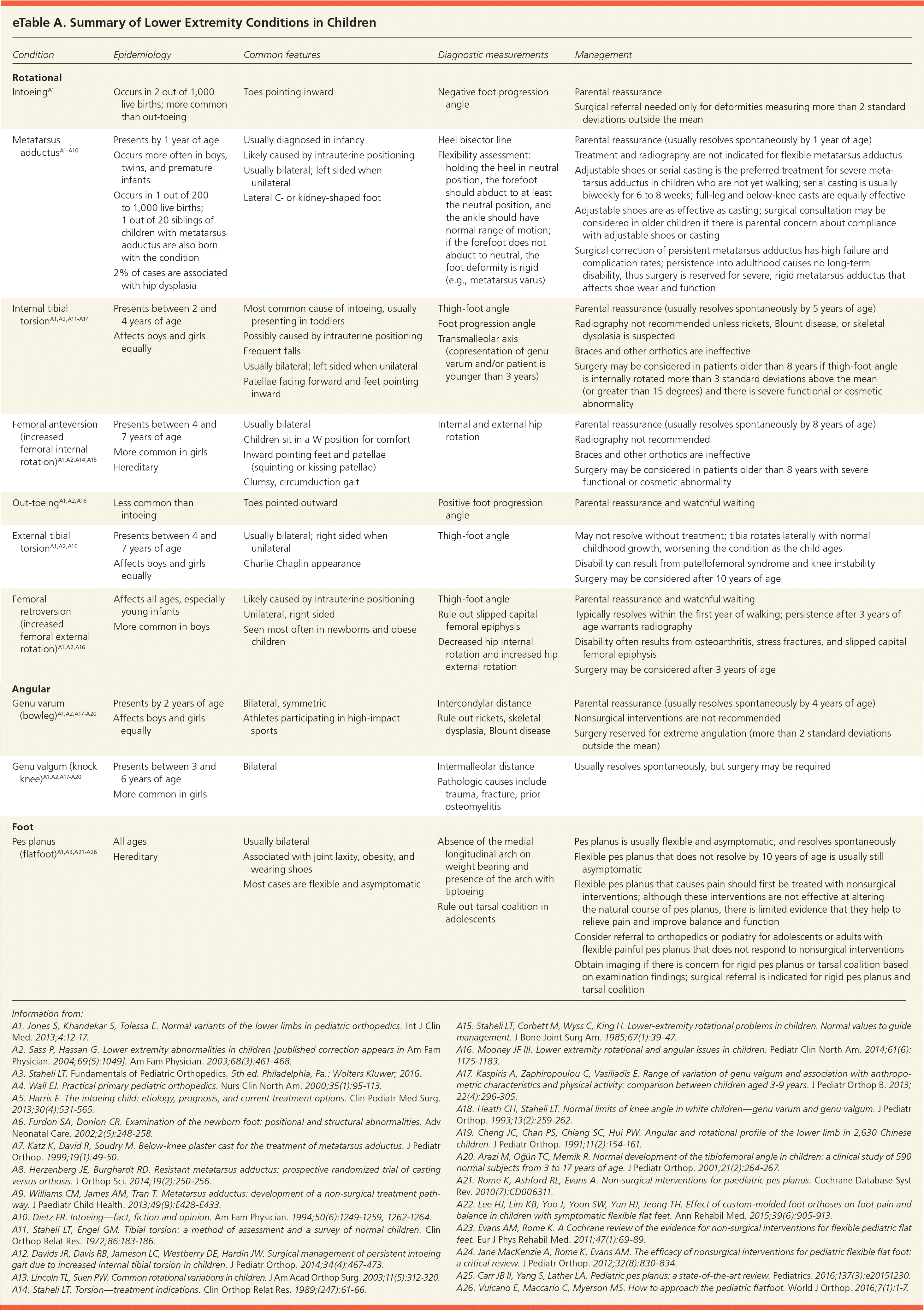
| Rotational |
| | | | |
| Presents by 1 year of age Occurs more often in boys, twins, and premature infants Occurs in 1 out of 200 to 1,000 live births; 1 out of 20 siblings of children with metatarsus adductus are also born with the condition 2% of cases are associated with hip dysplasia
| Usually diagnosed in infancy Likely caused by intrauterine positioning Usually bilateral; left sided when unilateral Lateral C- or kidney-shaped foot
| Heel bisector line Flexibility assessment: holding the heel in neutral position, the forefoot should abduct to at least the neutral position, and the ankle should have normal range of motion; if the forefoot does not abduct to neutral, the foot deformity is rigid (e.g., metatarsus varus)
| Parental reassurance (usually resolves spontaneously by 1 year of age) Treatment and radiography are not indicated for flexible metatarsus adductus Adjustable shoes or serial casting is the preferred treatment for severe metatarsus adductus in children who are not yet walking; serial casting is usually biweekly for 6 to 8 weeks; full-leg and below-knee casts are equally effective Adjustable shoes are as effective as casting; surgical consultation may be considered in older children if there is parental concern about compliance with adjustable shoes or casting Surgical correction of persistent metatarsus adductus has high failure and complication rates; persistence into adulthood causes no long-term disability, thus surgery is reserved for severe, rigid metatarsus adductus that affects shoe wear and function
|
| | Most common cause of intoeing, usually presenting in toddlers Possibly caused by intrauterine positioning Frequent falls Usually bilateral; left sided when unilateral Patellae facing forward and feet pointing inward
| | Parental reassurance (usually resolves spontaneously by 5 years of age) Radiography not recommended unless rickets, Blount disease, or skeletal dysplasia is suspected Braces and other orthotics are ineffective Surgery may be considered in patients older than 8 years if thigh-foot angle is internally rotated more than 3 standard deviations above the mean (or greater than 15 degrees) and there is severe functional or cosmetic abnormality
|
| | Usually bilateral Children sit in a W position for comfort Inward pointing feet and patellae (squinting or kissing patellae) Clumsy, circumduction gait
| | Parental reassurance (usually resolves spontaneously by 8 years of age) Radiography not recommended Braces and other orthotics are ineffective Surgery may be considered in patients older than 8 years with severe functional or cosmetic abnormality
|
| | | | |
| | | | May not resolve without treatment; tibia rotates laterally with normal childhood growth, worsening the condition as the child ages Disability can result from patellofemoral syndrome and knee instability Surgery may be considered after 10 years of age
|
| | | | Parental reassurance and watchful waiting Typically resolves within the first year of walking; persistence after 3 years of age warrants radiography Disability often results from osteoarthritis, stress fractures, and slipped capital femoral epiphysis Surgery may be considered after 3 years of age
|
| Angular |
| | | | Parental reassurance (usually resolves spontaneously by 4 years of age) Nonsurgical interventions are not recommended Surgery reserved for extreme angulation (more than 2 standard deviations outside the mean)
|
| | | | |
| Foot |
| | Usually bilateral Associated with joint laxity, obesity, and wearing shoes Most cases are flexible and asymptomatic
| | Pes planus is usually flexible and asymptomatic, and resolves spontaneously Flexible pes planus that does not resolve by 10 years of age is usually still asymptomatic Flexible pes planus that causes pain should first be treated with nonsurgical interventions; although these interventions are not effective at altering the natural course of pes planus, there is limited evidence that they help to relieve pain and improve balance and function Consider referral to orthopedics or podiatry for adolescents or adults with flexible painful pes planus that does not respond to nonsurgical interventions Obtain imaging if there is concern for rigid pes planus or tarsal coalition based on examination findings; surgical referral is indicated for rigid pes planus and tarsal coalition
|