Am Fam Physician. 2017;96(4):255-256
Author disclosure: No relevant financial affiliations.

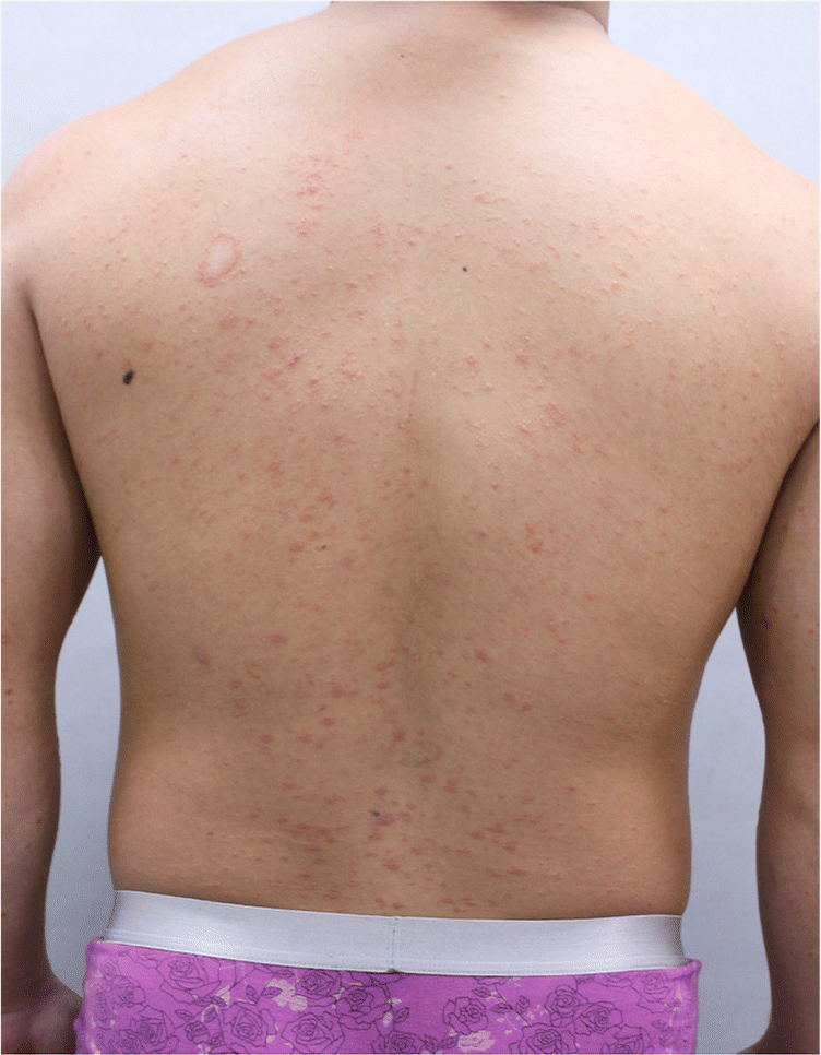
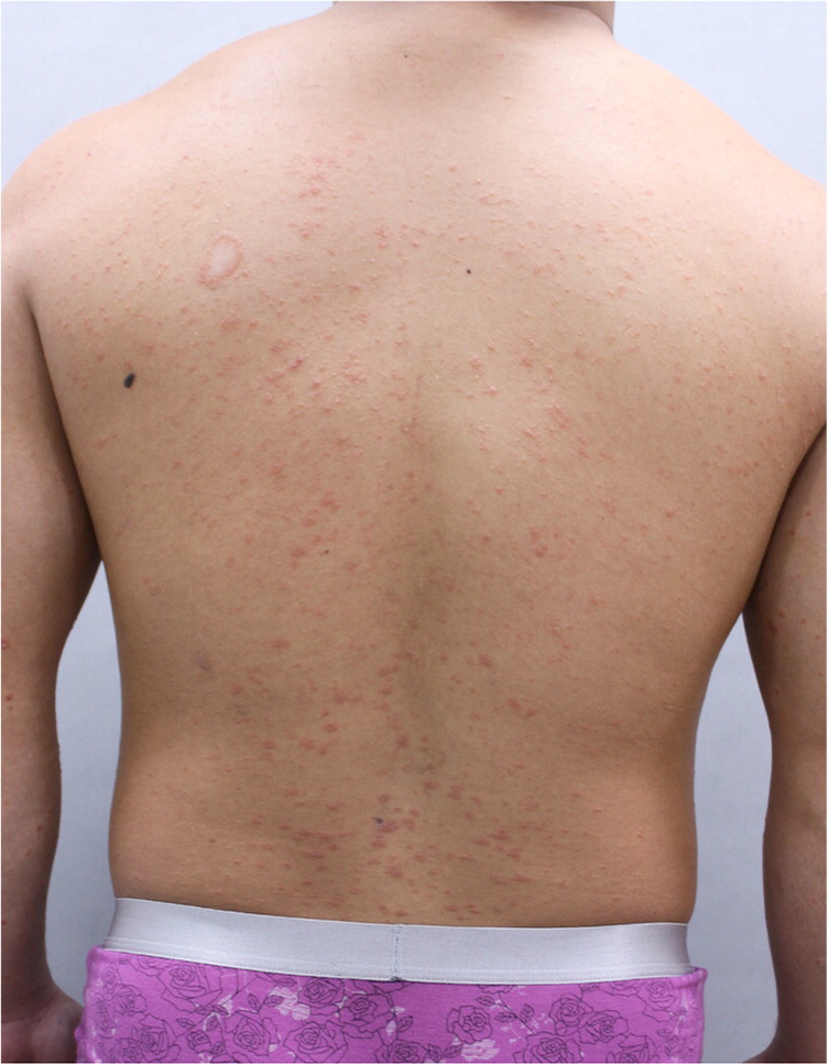
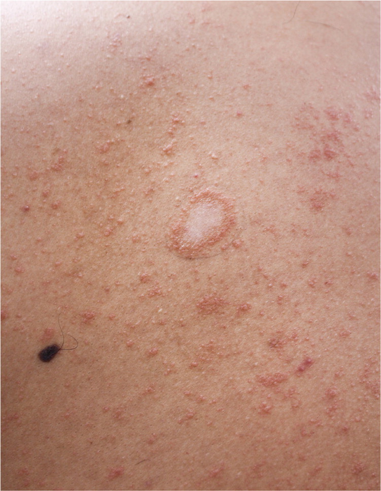
A 24-year-old man presented with a three-week history of a mildly pruritic, spreading rash. Before the generalized skin eruption, there was a single plaque on his back. Fatigue and upper respiratory symptoms preceded the appearance of the rash. There was no history of trauma to the area, including friction or rubbing. He had not recently used any topical agents or new medications.


Question
Discussion
The answer is C: pityriasis rosea. Typically, pityriasis rosea begins with a single erythematous, scaly patch on the trunk (herald patch). The herald patch appears one to 20 days before the generalized rash of pityriasis rosea. It is an oval, salmon-colored or red plaque with a scale trailing just inside the edge of the lesion like a collarette. The herald patch is usually 1.5 to 5 cm in size.
The generalized rash includes numerous smaller (1 cm), scaly, pink patches that develop on the trunk along the lines of skin cleavage. This has often been described as a Christmas tree pattern because skin cleavage lines run diagonally on the back. Pityriasis rosea lasts an average of eight to 12 weeks, although longer and shorter courses have been reported. The cause of pityriasis rosea is unknown, but there is some evidence of an infectious etiology. Pityriasis rosea resolves without treatment in one to three months.1,2
Granuloma annulare is a noninfectious granulomatous skin condition that can present as indurated and scaly dermal papules and annular plaques. Localized granuloma annulare is characterized by flesh-colored to violaceous lesions up to 5 cm in diameter. The pathogenesis of granuloma annulare is unknown, but it is thought to be a cell-mediated hypersensitivity reaction. Treatment is divided into localized skin-directed therapies and systemic immunomodulatory or immunosuppressive therapies.3,4
Guttate psoriasis is characterized by the acute onset of erythematous plaques or papules, often with a fine scale, that are generally less than 1 cm in size. They usually occur over the trunk and extremities in a centripetal pattern. The characteristic lesions appear as monomorphic droplets at the same stage of evolution. Guttate psoriasis often affects children and adolescents following a streptococcal infection (e.g., Streptococcus pyogenes) or an upper respiratory tract infection. It usually resolves without treatment within several weeks to months, but topical steroids can be effective.5
Tinea corporis is a superficial fungal infection of the skin. It presents as well-demarcated, erythematous papules or plaques that gradually enlarge over time. Potassium hydroxide microscopic testing can confirm the diagnosis. Topical anti-fungal medications are effective in treating localized lesions.3
| Condition | Characteristics |
|---|---|
| Granuloma annulare | Indurated, nonscaly, flesh-colored annular plaques and papules; usually on the extremities |
| Guttate psoriasis | Erythematous drop-like plaques, usually with a fine scale, over the trunk and extremities in a centripetal pattern |
| Pityriasis rosea | Oval, salmon-colored or red herald patch with a scale trailing just inside the edge of the lesion like a collarette; the generalized rash that follows includes numerous smaller, scaly, pink patches that develop on the trunk along the lines of skin cleavage |
| Tinea corporis | Well-demarcated, erythematous, enlarging papules or plaques |
