A 43-year-old man presented with a painful rash on the chest and back. The rash started five days earlier on the left side of the chest and then spread to the left side of the back. The pain was burning in nature. He had no history of a similar rash. He had no exposure to chemicals or physical agents. He had diabetes mellitus, hypertension, and dyslipidemia.
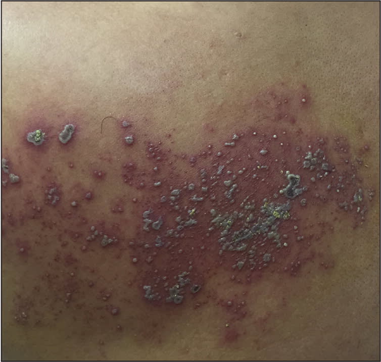
On physical examination, the patient was afebrile. Clusters of vesicles with some pustules and crusting (Figures 1 and 2) were noted on the back and chest. The rest of the body was not affected.
Figure 1.
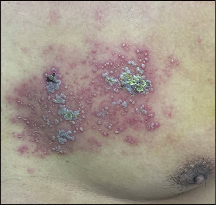
Figure 2.
Question
Based on the patient's history and physical examination findings, which one of the following is the most likely diagnosis?
A. Bullous impetigo.
B. Contact dermatitis.
C. Herpes simplex virus infection.
D. Herpes zoster.
E. Varicella.
Discussion
The answer is D: herpes zoster. Herpes zoster is characterized by a unilateral, painful vesicular rash with a dermatomal distribution. It is caused by reactivation of the varicella zoster virus.1 Herpes zoster most commonly affects dermatomes T1 to L2 and the first branch of the trigeminal nerve.1 In this case, the patient had a painful vesicular rash distributed mainly at the T2 to T4 dermatomes. There are four stages of a varicella zoster outbreak: erythematous, vesicular, pustular, and ulcerative.1 Occasionally, crusting develops after one week.2 The pain, which is caused by acute neuritis, can be described as a burning sensation and a deep, prickling, or lancinating pain.2 Reduced immunity states, such as advanced age and immunosuppression (e.g., malignancy, diabetes, human immunodeficiency virus infection), are risk factors for herpes zoster.2
Bullous impetigo is a result of superficial Staphylococcus aureus or group A Streptococcus skin infections.2 It usually arises from a primary infection in minor superficial skin breaks or secondary infection of preexisting dermatoses.2 The vesicles have sharp margins with no surrounding erythema.3 Bullous impetigo is more common in moist, intertriginous areas, such as diaper areas, axillae, and neck folds.2,3
Contact dermatitis is an acute or chronic inflammatory reaction to substances that come into contact with the skin.2 There must be a triggering agent to cause the reaction. Lesions vary and can include erythema, vesicles, blisters, erosions, and crusts.2
Herpes simplex virus infection usually occurs at mucocutaneous areas, such as the orolabial area and genitalia.4 The lesions are clusters of painful vesicles on an erythematous base, which can evolve into pustules, erosions, and ulcerations.2 Widespread cutaneous herpes simplex virus infection can occur on the trunk in patients with atopic dermatitis or an immunocompromising condition.2 The lesions will be generalized and disseminated rather than grouped.2
Varicella, or chicken pox, is characterized by crops of pruritic rash with lesions in different stages of development (macules, papules, vesicles, pustules, and crusted scabs).2,5 The lesions usually begin on the head and spread to the trunk and extremities in a bilateral distribution.2 The rash is pruritic rather than painful.3
Summary Table
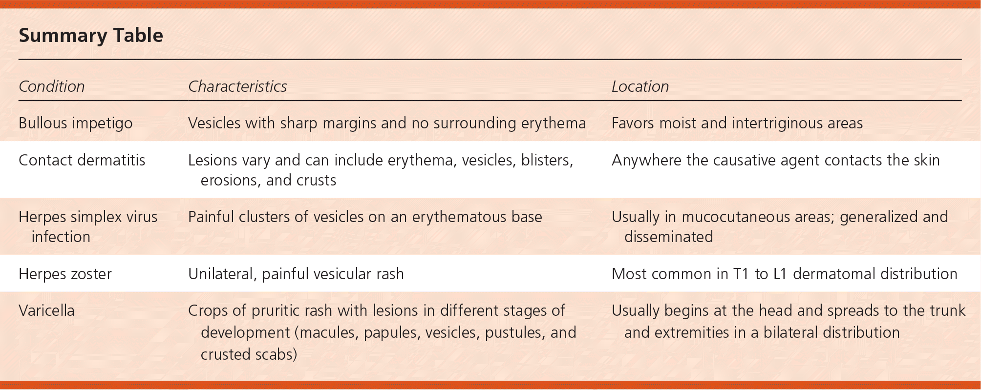
| Condition | Characteristics | Location |
|---|---|---|
| Bullous impetigo | Vesicles with sharp margins and no surrounding erythema | Favors moist and intertriginous areas |
| Contact dermatitis | Lesions vary and can include erythema, vesicles, blisters, erosions, and crusts | Anywhere the causative agent contacts the skin |
| Herpes simplex virus infection | Painful clusters of vesicles on an erythematous base | Usually in mucocutaneous areas; generalized and disseminated |
| Herpes zoster | Unilateral, painful vesicular rash | Most common in T1 to L1 dermatomal distribution |
| Varicella | Crops of pruritic rash with lesions in different stages of development (macules, papules, vesicles, pustules, and crusted scabs) | Usually begins at the head and spreads to the trunk and extremities in a bilateral distribution |
