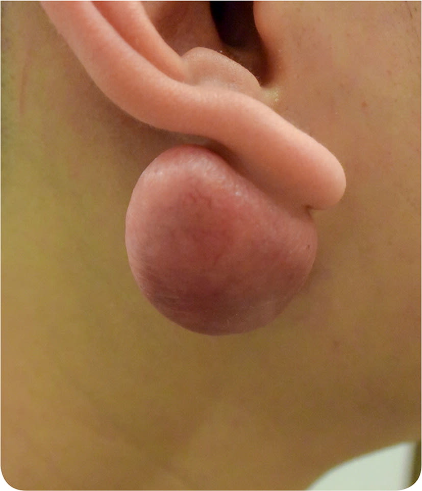
A 23-year-old man presented with a painless mass behind his right earlobe that had been present for one year (Figure 1). He initially noticed a flesh-colored, pinpoint growth on both sides of his earlobe after wearing a fake hoop earring that compressed his earlobe. Although the changes on the front of the earlobe resolved, the posterior mass gradually increased in size. There was no pain, itching, or bleeding.
On physical examination, there was a mass measuring 1.5 × 2 cm on the posterior aspect of his right earlobe. There were no skin changes, although small blood vessels could be seen extending into the lesion. On palpation, the mass was slightly firm, nontender, and nonpulsatile, and it did not transilluminate.
FIGURE 1
Question
Based on the patient's history and physical examination findings, which one of the following is the most likely diagnosis?
A. Acrochordon.
B. Epidermal inclusion cyst.
C. Hypertrophic scar.
D. Keloid.
Discussion
The answer is D: keloid. The earlobe is a common site for keloids, which are benign dense growths of disorganized collagen fibers that occur in response to skin trauma. Keloids can extend beyond the border of the initial injury, have delayed appearance, and grow indefinitely, and they often regrow after removal.1 Auricular keloids occur with 2.5% of all ear piercings.2 Keloid removal can be challenging because of the potential for disfigurement or further scarring. Keloids most often affect persons 10 to 30 years of age.2
Treatment options are varied and include excision, intralesional corticosteroids, excision combined with corticosteroid injection, cryotherapy, silicone sheeting, pressure dressings, immunosuppressive or antitumor agents, pulsed dye laser treatment, and radiation therapy. Rate of recurrence for excisional removal varies widely, with case studies reporting rates of 50% to 100%.1 Recurrence is reduced to 10% to 50% when excision is augmented with intralesional corticosteroids at the time of removal or afterward.1 Cryotherapy can be effective on smaller lesions or in conjunction with corticosteroid injections without excision. Pressure dressings can be used after removal of auricular keloids; however, they can be painful and difficult to apply.1
Acrochordons, or skin tags, present as soft, pedunculated, or sessile lesions at the site of skin irritation or trauma. Skin tags are usually flesh colored but may be slightly darker.3,4
Epidermal inclusion cysts are benign overgrowths of keratinous material that are derived from epidermoid cells and surrounded by stratified squamous epithelium in the dermal layer of the skin. They are one of the most common skin lesions. Inclusion cysts are flesh colored, have a central punctum, and are mobile within the dermis. These cysts can develop spontaneously, in response to trauma, or from a comedone. They vary in size from a few millimeters to several centimeters. They can remain unchanged for years, but they may become inflamed or infected over time.3
Hypertrophic scars are fibrous scars that occur in response to skin trauma and are similar to keloids. However, they stay within the borders of the wound, tend to develop more quickly after trauma, and regress spontaneously, usually within one year of injury.5
SUMMARY TABLE
| Condition | Characteristics |
|---|---|
| Acrochordon | Soft, pedunculated or sessile lesions at the sites of skin irritation or trauma; flesh colored or slightly darker |
| Epidermal inclusion cyst | Flesh-colored lesion with central punctum; firm but mobile within the dermis; may be painful if infected or inflamed; contents are heterogeneous and can be drained |
| Hypertrophic scar | Fibrous scar at site of skin trauma; occurs within one month of injury and does not exceed borders of the original wound |
| Keloid | Benign dense fibrous growth at site of skin trauma; can extend beyond the border of the initial injury, have delayed appearance, and grow indefinitely; often regrow after removal |
