A 41-year-old man presented with foot pain, which had been present for three months. He also had lesions on his elbows and knees that had been present for about five years. The patient's history included tobacco use, newly diagnosed hypertension, and a recent transient ischemic attack. Before the transient ischemic attack, he was not seeing a physician regularly.
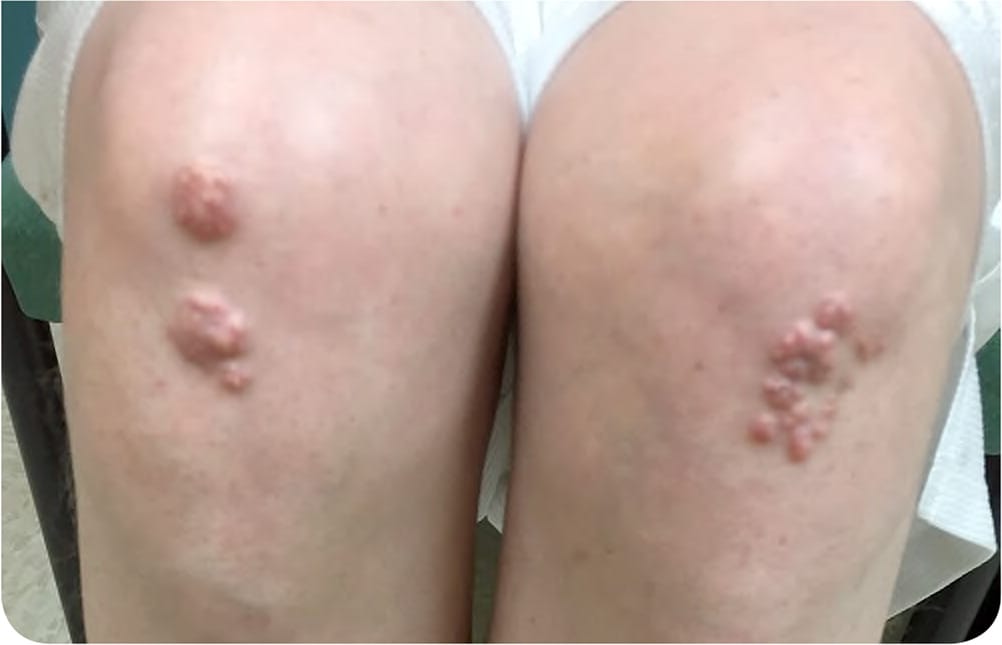
Physical examination revealed nontender, red-yellow papules scattered across the extensor surfaces of the elbows and knees (Figure 1 and Figure 2). They were approximately 3 mm to 1 cm in size.
FIGURE 1
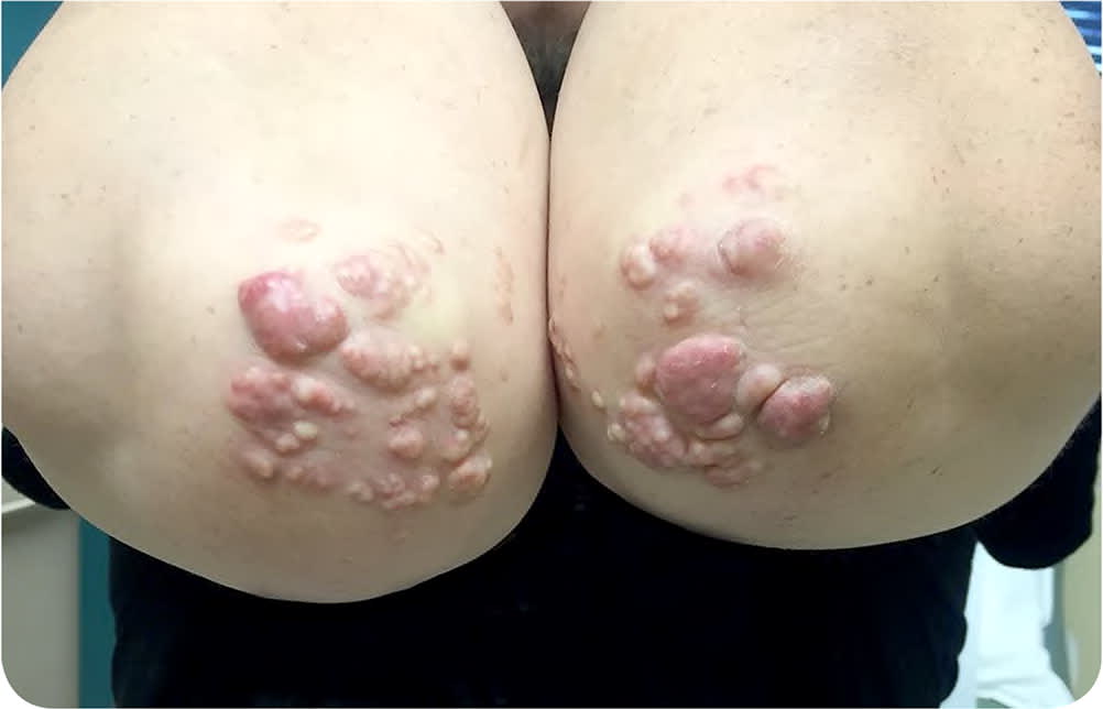
FIGURE 2
Question
Based on the patient's history and physical examination findings, which one of the following is the most likely diagnosis?
A. Gouty tophi.
B. Granulomatosis with polyangiitis.
C. Rheumatoid nodules.
D. Sarcoidosis.
E. Xanthomas.
Discussion
The answer is E: xanthomas. Xanthomas are nontender lesions consisting of abnormal lipid deposition and foam cells.1 They are characterized by accumulations of lipid-laden macrophages and can develop in the setting of altered lipid metabolism, although they occasionally occur without underlying metabolic effects.1,2 Xanthomas do not represent a disease; rather, they are a sign of a variety of lipoprotein disorders.1 One major clinical feature of xanthomas is the yellow to red hue. Morphology can vary from macules and papules to plaques and nodules.3 They can develop on the skin and tendons.
Xanthomas are classified into four categories: eruptive, planar, tendinous, and tuberoeruptive or tuberous. Eruptive xanthomas are yellow-red papules that appear suddenly in crops on extensor surfaces of the extremities and the buttocks. Planar xanthomas are yellow macules, papules, or plaques on the upper eyelids, wrists, palms, and intertriginous areas. Tendinous xanthomas are firm nodules that develop subcutaneously in fasciae, ligaments, and tendons, often in extensor surfaces.1 Tuberoeruptive and tuberous xanthomas are clinically related and are thought to be on a continuum. They often present as pink-yellow papules or nodules on extensor surfaces, specifically the elbows and knees. Tuberous lesions are the larger of the two, exceeding 3 cm in diameter. Tuberoeruptive and tuberous xanthomas can occur with hypercholesterolemic states such as dysbetalipoproteinemia (Frederickson type III) and familial hypercholesterolemia (Frederickson type II).3
Gouty tophi are typically associated with poorly controlled gout. The tophi result from deposition of uric acid crystals in the soft tissue, including articular structures, tendons, or bursae. Gouty tophi are nontender, may be yellowish white in color, and are commonly seen in toes, fingers, ears, and elbows.1
Granulomatosis with polyangiitis, or Wegener granulomatosis, is an autoimmune disease with necrotizing granulomatous inflammation. It can present as palpable purpura, papules, vesicles, subcutaneous nodules, plaques, and ulcers, typically with upper airway involvement and nonspecific constitutional symptoms such as fever, weight loss, and loss of appetite. The lesions are more common on the lower extremities.
Rheumatoid nodules are nontender and typically occur along the entire extensor surfaces of the forearms, although they are also common on the elbows. Patients with rheumatoid nodules usually have high titers of rheumatoid factor and rheumatoid arthritis.1
Sarcoidosis is a granulomatous disease that may present as painful maculopapular lesions on the head and neck or plaques on the extensor surface of the lower extremities. The lesions often are not symmetric or bilateral. Pulmonary involvement occurs in most patients with sarcoidosis.1
SUMMARY TABLE
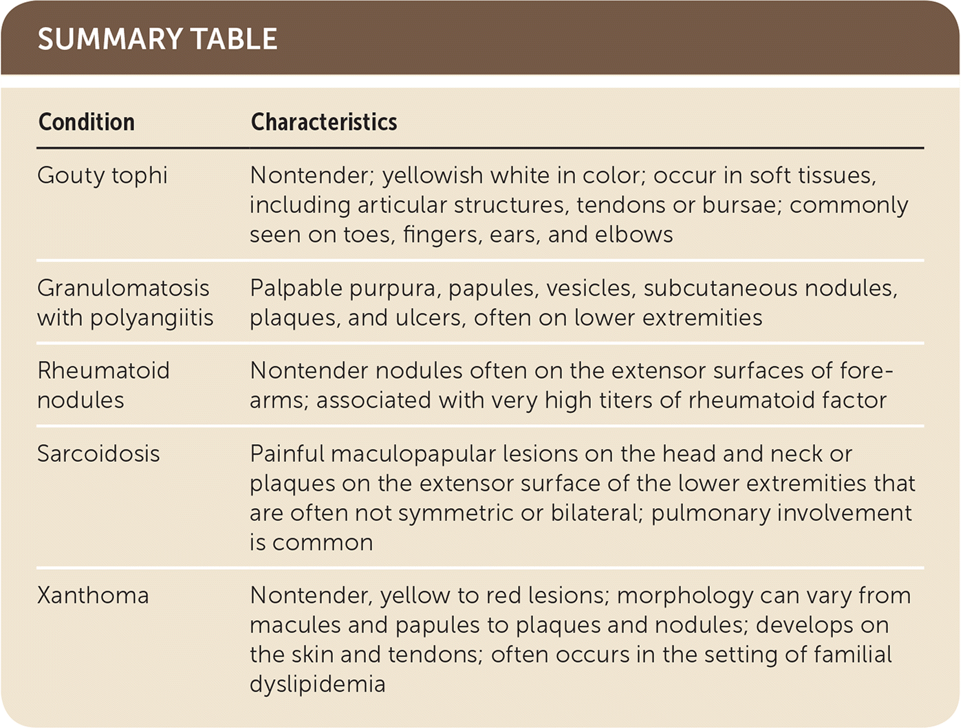
| Condition | Characteristics |
|---|---|
| Gouty tophi | Nontender; yellowish white in color; occur in soft tissues, including articular structures, tendons or bursae; commonly seen on toes, fingers, ears, and elbows |
| Granulomatosis with polyangiitis | Palpable purpura, papules, vesicles, subcutaneous nodules, plaques, and ulcers, often on lower extremities |
| Rheumatoid nodules | Nontender nodules often on the extensor surfaces of forearms; associated with very high titers of rheumatoid factor |
| Sarcoidosis | Painful maculopapular lesions on the head and neck or plaques on the extensor surface of the lower extremities that are often not symmetric or bilateral; pulmonary involvement is common |
| Xanthoma | Nontender, yellow to red lesions; morphology can vary from macules and papules to plaques and nodules; develops on the skin and tendons; often occurs in the setting of familial dyslipidemia |
