A 16-year-old girl presented with asymptomatic, white lesions on the hands and lower extremities. The lesions were first noted five years earlier and had slowly increased in size. There was no family history of a similar skin disorder or autoimmune disease.
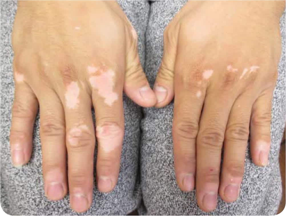
Physical examination revealed well-demarcated, chalk-white macules and patches on the dorsum of the hands (Figure 1) and extensor aspects of the legs.
FIGURE 1
Question
Based on the patient's history and physical examination findings, which one of the following is the most likely diagnosis?
A. Hypomelanosis of Ito.
B. Idiopathic guttate hypomelanosis.
C. Pityriasis alba.
D. Tinea versicolor.
E. Vitiligo.
Discussion
The answer is E: vitiligo. Vitiligo is characterized by asymptomatic, acquired, amelanotic macules and patches that are milky or chalk-white in color.1 Lesions are well demarcated, often symmetrical, and show homogenous depigmentation. The most common location is the face, followed by the neck, limbs, and trunk. The onset of vitiligo occurs before 10 years of age in 25% of patients, before 20 years in 50% of patients, and before 40 years in 95% of patients.1
In most patients, the disease is slowly progressive, with the appearance of new lesions or enlargement of existing lesions. Vitiligo is more common in those who have a family member with the condition. The cause of vitiligo is unclear, but the patches of depigmented skin are due to the loss of melanocytes.
Hypomelanosis of Ito is characterized by patterned, hypopigmented macules in well-demarcated streaks, stripes, whorls, and patches. It usually involves more than two body segments, most commonly the trunk and proximal extremities. The lesions often present at birth or in the first two years of life and tend to fade in adulthood. Chromosomal abnormalities are found in approximately 50% of cases.2 About 30% of affected patients have extracutaneous anomalies such as short stature, digital deformity, kyphoscoliosis, psychomotor retardation, hemimegalencephaly, hypotonia, autism, epilepsy, scleral melanocytosis, nystagmus, and strabismus.2
Idiopathic guttate hypomelanosis is characterized by multiple asymptomatic, discrete, well-circumscribed, smooth, porcelain-white macules with round or angular borders. Sites of predilection typically include areas with repeated sun exposure, such as the forearms and shins. The condition is more common after 30 years of age. The lesions tend to persist and may slowly enlarge.
Pityriasis alba is characterized by asymptomatic, hypopigmented, round or oval macules and patches with fine, loosely adherent scales and indistinct borders.3 The most common site is the face, especially the malar areas. The condition occurs predominantly in children three to 16 years of age, typically in those with darker skin tones. It is usually self-limited.3
Tinea versicolor is a fungal infection characterized by asymptomatic, hypo- or hyperpigmented macules. It most commonly affects areas of skin that are rich in sebum production, such as the trunk, upper arms, and neck. Fair-skinned patients tend to have red to brown lesions. Patients with darker skin tend to have hypopigmented lesions. The lesions are covered with a fine scale. The condition is most common among adolescents and young adults. Without antifungal treatment, the disease tends to persist and may slowly enlarge.
SUMMARY TABL E
| Condition | Onset | Location | Prognosis | Characteristics |
|---|---|---|---|---|
| Hypomelanosis of Ito | Birth or first two years of life | Trunk and proximal extremities | Tends to fade in adulthood | Patterned, hypopigmented macules in well-demarcated streaks, stripes, whorls, and patches |
| Idiopathic guttate hypomelanosis | More common after 30 years of age | Areas with repeated sun exposure, such as forearms and shins | Tends to persist and may slowly enlarge | Asymptomatic, discrete, well-circumscribed, smooth, porcelain-white macules with round or angular borders |
| Pityriasis alba | Three to 16 years of age | Face | Spontaneous resolution in two to three years | Asymptomatic, hypopigmented, round or oval macules and patches with fine, loosely adherent scales and indistinct borders |
| Tinea versicolor | Adolescents and young adults | Trunk, upper arms, and neck | Tends to persist and may slowly enlarge | Asymptomatic, hypo- or hyperpigmented macules that are covered with a fine scale |
| Vitiligo | Usually before 40 years of age | Face, neck, limbs, trunk | Slowly progressive | Asymptomatic, acquired, well-demarcated, milky or chalk-white macules or patches |
