A 73-year-old woman presented with asymmetrical pupils. She had no loss of vision, double vision, eye pain, light sensitivity, or abnormal sweating. The patient had a history of bilateral cataract surgery, repair of right retinal detachment, and repair of the right orbital bone after a traumatic event. She had a 30-pack-year history of smoking. Although the patient had several medical conditions, none involved the eye or use of ophthalmic medications.
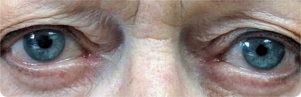
On physical examination, the external eyelids were normal in appearance with no findings of ptosis. The sclerae were clear bilaterally, with an approximately 1-mm difference in pupillary size observed in light and darkness (Figure 1). Pupils were round and reactive to light. Cranial nerves were intact with no abnormality noted on confrontation of visual fields.
Recent lung cancer screening with low-dose computed tomography revealed multiple lung nodules; however, none were in the apical region. Additionally, magnetic resonance imaging with and without contrast media was obtained during a prior emergency department visit as part of a stroke workup. It showed chronic microvascular ischemic disease with no evidence of a mass.
FIGURE 1
Question
Based on the patient's history and physical examination findings, which one of the following is the most likely diagnosis?
- A. Horner syndrome.
- B. Physiologic anisocoria.
- C. Third nerve palsy.
- D. Traumatic mydriasis.
Discussion
The answer is B: physiologic anisocoria. Physiologic anisocoria is the most common etiology of uneven pupil size, occurring in approximately 20% to 24% of the U.S. population. It is a benign condition, and no treatment is required.1–3
The diagnosis of physiologic anisocoria is clinical, based on history and physical examination findings. Reviewing old photos or driver's licenses of the patient can assist in establishing the diagnosis because physiologic anisocoria is a persistent and long-standing condition.3,4 The anisocoria is usually equal in light and darkness.3
Horner syndrome presents as the classic triad of unilateral miosis, ptosis, and anhidrosis.5 The anisocoria is greater in darkness than in light, and the abnormal pupil is typically the smaller pupil.3 Evaluation includes urgent imaging, including magnetic resonance imaging and magnetic resonance angiography of the neck, to rule out carotid dissection. Images should extend to the hypothalamus and upper chest to identify lesion location.3,6
There are multiple etiologies for third nerve palsy, including trauma, cerebrovascular disease, and inflammatory disorders. Signs and symptoms include diplopia, ptosis, and impaired extraocular movements. The anisocoria is greater in the light, and the abnormal pupil is typically the larger pupil. Similar to Horner syndrome, third nerve palsy requires urgent neuroimaging, including computed tomography angiography, to evaluate for presence of an intracranial aneurysm.6
Traumatic mydriasis is caused by ocular injury to the pupillary sphincter muscle.3 The anisocoria is more prominent with bright light because the pupil is unable to constrict as a result of the injury.
SUMMARY TABLE
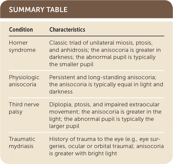
| Condition | Characteristics |
|---|---|
| Horner syndrome | Classic triad of unilateral miosis, ptosis, and anhidrosis; the anisocoria is greater in darkness; the abnormal pupil is typically the smaller pupil |
| Physiologic anisocoria | Persistent and long-standing anisocoria; the anisocoria is typically equal in light and darkness |
| Third nerve palsy | Diplopia, ptosis, and impaired extraocular movement; the anisocoria is greater in the light; the abnormal pupil is typically the larger pupil |
| Traumatic mydriasis | History of trauma to the eye (e.g., eye surgeries, ocular or orbital trauma); anisocoria is greater with bright light |
