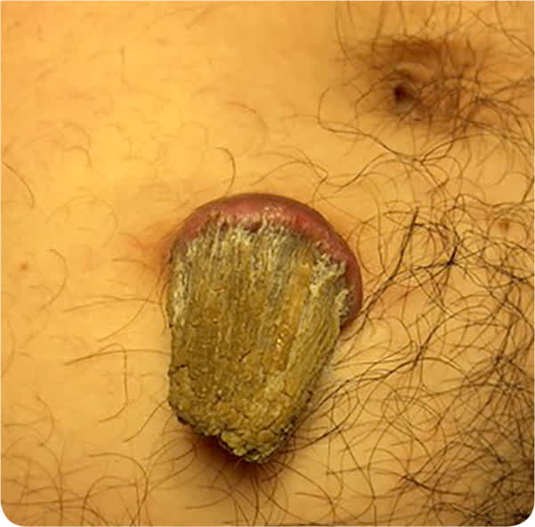
A 36-year-old man presented with a growth on his right lower abdomen. The nodule was initially small but had grown over the previous three years. The patient was unsure if it began as a wound or abscess. It was drained once in the emergency department. The area was not painful or tender, but the lesion was intermittently pruritic because it was located near his belt buckle. He had no significant medical history.
On physical examination, the patient was in no distress and had normal vital signs. The base of the nodule was about 2 cm in size and was not erythematous, warm to the touch, or macerated (Figure 1). The base of the nodule was hyperkeratotic, but no fluctuance or abscess was palpable underneath.
FIGURE 1
Question
Based on the patient's history and physical examination findings, which one of the following is the most likely diagnosis?
A. Giant molluscum contagiosum.
B. Kaposi sarcoma.
C. Keratoacanthoma.
D. Merkel cell carcinoma.
E. Nodular basal cell carcinoma.
Discussion
The answer is C: keratoacanthoma. A keratoacanthoma typically presents as a skin nodule with a centralized keratin filling. It most commonly develops on sun-exposed skin.1 The pathogenesis is not known. Keratoacanthomas are often diagnosed clinically. They have a rapid proliferative stage for the first six to eight weeks. Growth may stop for several months and then spontaneously regress.2 Because it is difficult to distinguish between keratoacanthoma and squamous cell carcinoma based on gross appearance, it is prudent to confirm the diagnosis with a biopsy.
An elliptical excision was performed for this patient's lesion. An incomplete removal can result in recurrence.3 Therefore, at the patient's follow-up appointment, we ensured that the lesion was healing well and that there were no signs of regrowth.
Giant molluscum contagiosum is a common benign viral infection caused by the poxvirus that affects the skin. It commonly occurs in young children, presenting as umbilicated papules on the face, arms, and legs, but can also affect patients who are immunocompromised. It is diagnosed visually. Although the papules are normally smaller than 5 mm and fewer than 20 in quantity, giant molluscum can grow to larger than 10 mm. The lesions can appear throughout the body.4
Kaposi sarcoma is a vascular tumor that affects the skin and internal organs. It is known for its association with human immunodeficiency virus infection but can also manifest with human herpesvirus 8 infection. Kaposi sarcoma presents as dark brown or reddish patches, papules, plaques, or deeper skin nodules. In the urogenital region, Kaposi sarcoma usually affects the penis.5
Merkel cell carcinoma is a rare, rapidly growing neuroendocrine tumor often found on the head or neck. It presents as a firm, erythematous nodule in patients with impaired immune systems, including older people, those with AIDS, or those taking immunosuppressive medications. A lymph node evaluation is required to diagnose Merkel cell carcinoma because of its aggressive nature and poor prognosis.6
Nodular basal cell carcinoma is the most common form of basal cell carcinomas. Basal cell carcinomas are slow-growing tumors that develop on areas of chronic sun exposure, including the face. They classically present as shiny, flesh-colored nodules with recurrent bleeding. The nodules can ulcerate; therefore, the borders appear to be rolled.7
SUMMARY TABLE
| Condition | Characteristics |
|---|---|
| Giant molluscum contagiosum | Umbilicated papules; typically small but can be larger than 10 mm |
| Kaposi sarcoma | Dark brown or reddish patches, papules, plaques, or nodules |
| Keratoacanthoma | Rapidly growing skin nodule with a centralized keratin filling |
| Merkel cell carcinoma | Rapidly growing, firm, erythematous nodule |
| Nodular basal cell carcinoma | Slow-growing, shiny, flesh-colored nodules with recurrent bleeding; nodules can ulcerate |
