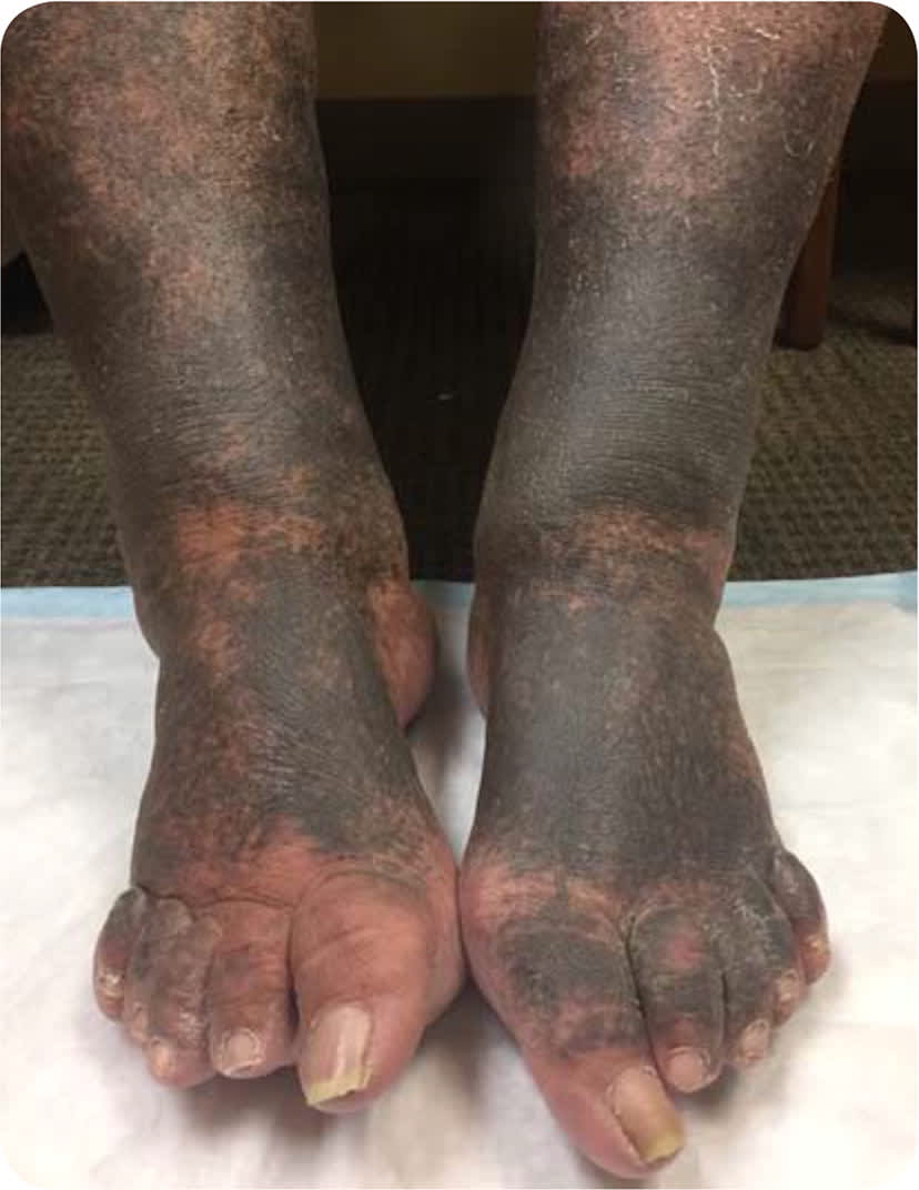
A 68-year-old obese man presented for chronic disease follow-up. The patient's only concern was discoloration in his lower extremities that had developed over the previous three weeks. No other areas were involved, and he had no pain or itching.
His medical history included diabetes mellitus, hypertension, hyperlipidemia, and obstructive sleep apnea. His medications included hydrochlorothiazide, metformin, glipizide (Glucotrol), minocycline (Minocin), and pravastatin (Pravachol). Six weeks before presentation, he had right hip arthroplasty with postoperative joint infection that required prolonged suppressive antibiotic treatment. His vital signs were normal on physical examination. A diffuse area of hyperpigmentation was present on the lower legs and feet. The rash was bilateral with dark hyperpigmentation (Figure 1). Lower extremity distal pulses were normal.
FIGURE 1
Question
Based on the patient's history and physical examination findings, which one of the following is the most likely diagnosis?
A. Addison disease.
B. Diabetic dermopathy.
C. Hemochromatosis.
D. Medication reaction.
E. Venous insufficiency.
Discussion
The answer is D: medication reaction. Cutaneous hyperpigmentation is a recognized adverse effect of long-term minocycline therapy. The skin discoloration is largely cosmetic and not associated with other adverse clinical effects.1
The hyperpigmentation results from deposition of insoluble minocycline-iron complexes and can be blue-gray or muddy brown in color. The discoloration can occur on the face, arms, shins, or legs. The incidence of minocycline-related hyperpigmentation is highest in patients receiving long-term therapy for chronic infection (54%) or rheumatoid arthritis (36%). The incidence in those receiving low-dose minocycline for acne vulgaris is low (2.4%).2 In general, the risk of hyperpigmentation increases with longer duration of use.1,2
Treatment for minocycline-induced hyperpigmentation can be difficult. Effective resolution of hyperpigmentation has been shown with use of Q-switched alexandrite laser irradiation; however, availability of this therapy is limited.3 Even with cessation of minocycline use, skin discoloration may persist indefinitely, and many patients choose to continue taking the medication. However, they should be counseled that additional body areas could be affected with continued use.
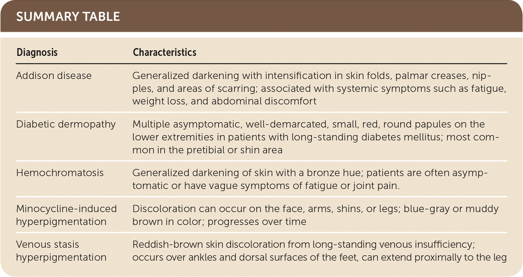
Addison disease, or adrenocortical insufficiency, is caused by the destruction or dysfunction of the adrenal cortex. It affects glucocorticoid and mineralocorticoid function. It is associated with systemic symptoms such as fatigue, weight loss, and abdominal discomfort. Addison disease can lead to generalized darkening with intensification in skin folds, palmar creases, nipples, and areas of scarring.2
Diabetic dermopathy occurs on the lower extremities in patients with long-standing diabetes. It typically presents as multiple asymptomatic, well-demarcated, small, red, round papules that heal and disappear with time, often leaving residual hypopigmentation. It is most common in the pretibial or shin area.4
Hyperpigmentation from hemochromatosis is generalized darkening of the skin with a bronze hue. It is often asymptomatic or causes vague symptoms of fatigue or joint pain. Serum ferritin and transferrin saturation are elevated.5
Venous stasis hyperpigmentation results from chronic venous drainage insufficiency in the lower extremity. The hyperpigmentation is preceded by chronic leg swelling and stasis dermatitis. The skin discoloration is typically reddish-brown and most prominent at the medial ankle with slow progression into the foot and lower leg.6
SUMMARY TABLE
| Diagnosis | Characteristics |
|---|---|
| Addison disease | Generalized darkening with intensification in skin folds, palmar creases, nipples, and areas of scarring; associated with systemic symptoms such as fatigue, weight loss, and abdominal discomfort |
| Diabetic dermopathy | Multiple asymptomatic, well-demarcated, small, red, round papules on the lower extremities in patients with long-standing diabetes mellitus; most common in the pretibial or shin area |
| Hemochromatosis | Generalized darkening of skin with a bronze hue; patients are often asymptomatic or have vague symptoms of fatigue or joint pain. |
| Minocycline-induced hyperpigmentation | Discoloration can occur on the face, arms, shins, or legs; blue-gray or muddy brown in color; progresses over time |
| Venous stasis hyperpigmentation | Reddish-brown skin discoloration from long-standing venous insufficiency; occurs over ankles and dorsal surfaces of the feet, can extend proximally to the leg |
