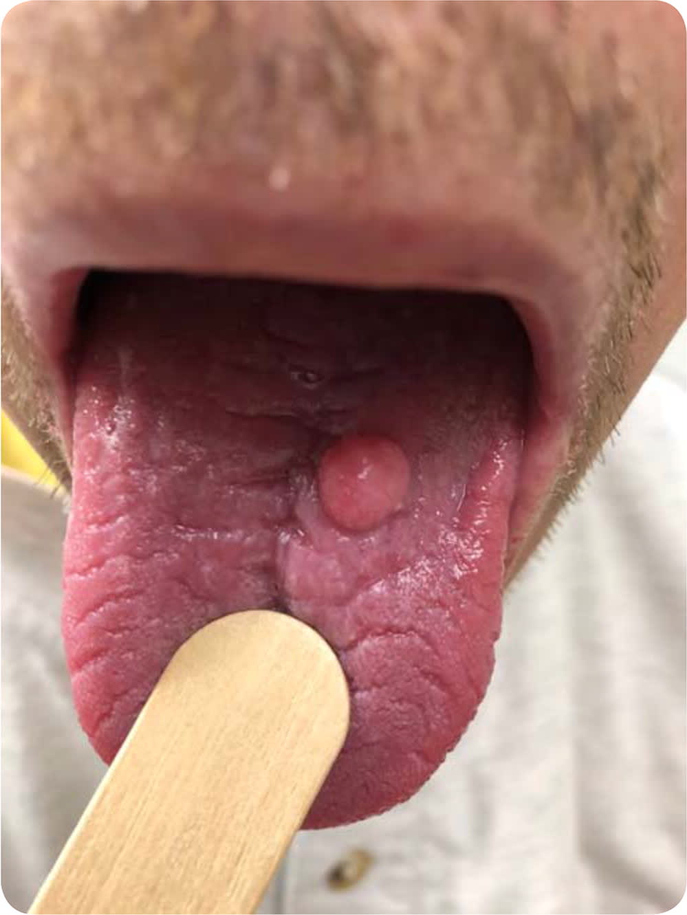
A 51-year-old man presented with a nodule on his tongue that had been growing slowly for three years. The nodule had become painful and interfered with his speech and oral intake. He did not recall any injury or trauma to his mouth. The lesion had never bled, ruptured, or ulcerated. His medical history was significant for current tobacco use, chronic hepatitis C, and past intravenous drug use.
Physical examination revealed an 8-mm, well-circumscribed, smooth, pink, firm nodule on the dorsal aspect of the tongue (Figure 1). A punch biopsy showed squamous proliferation with papillary surface projections and virally altered cells.
FIGURE 1
Question
Based on the patient's history, physical examination, and biopsy findings, which one of the following is the most likely diagnosis?
A. Condyloma acuminatum.
B. Irritation fibroma.
C. Oral squamous papilloma.
D. Pyogenic granuloma.
E. Squamous cell carcinoma.
Discussion
The answer is A: condyloma acuminatum, a highly contagious skin lesion caused by infection with human papillomavirus (HPV) type 2, 6, 11, 16, or 18.1 Although condyloma acuminatum is usually caused by low-risk HPV types, infection with the high-risk types 16 and 18 has been linked with anogenital, head, and neck cancers.2 Condyloma acuminatum typically occurs in the anogenital region but can involve the oral cavity via oral-genital contact or autoinoculation.3 Oral condyloma acuminatum is usually asymptomatic and can appear as single or multiple pedunculated, smooth, or dome-shaped papules or plaques.1 Histopathologic features include broad hyperplastic papillary projections with cells demonstrating viral changes, such as koilocytosis, parakeratosis, and increased nuclear atypia.4 Condyloma acuminatum is treated with surgical excision or cauterization.1
Irritation fibromas are caused by chronic irritation of the oral mucosa and appear along the bite line as a single smooth, dome-shaped papule.5 Because this benign change can be difficult to differentiate from malignant neoplasms, excisional biopsy is recommended.5 Histopathologic findings of irritation fibromas include a mass of fibrous tissue covered by normal squamous mucosa.4
Oral squamous papillomas are the most common benign oral neoplasms.5 They can occur anywhere in the mouth but have a predilection for the ventral surface of the tongue. They usually appear as a single exophytic growth.5 Squamous papillomas are typically related to low-risk HPV types 6 and 11.5 These lesions can appear similar to condyloma acuminatum but are usually smaller and not clustered. Histopathology shows squamous epithelium arrayed in finger-like projections with surrounding hyperkeratosis.4 Oral squamous papillomas are treated with surgical excision.5
Pyogenic granulomas are benign, vascular tumors that can occur anywhere on the skin or mucous membranes. They appear as a single rapidly growing, friable, red papule usually on the lip or gingival mucosa. They may be pedunculated or sessile.6 Histopathology demonstrates a lobular pattern of vascular proliferation with inflammation and edema.4 The cause is unknown, but trauma and irritation are possible triggers.6 Pyogenic granulomas are treated with surgical excision followed by electrodesiccation to control bleeding.6
Squamous cell carcinoma is the most common cancer of the oral cavity. Major risk factors include tobacco and alcohol use.2 Oral squamous cell carcinoma can present as thickened white or red patches, ulcerations, or papules that are usually localized to the lateral tongue.7 Shave, punch, or excisional biopsy is recommended for diagnosis. Histopathology can consist of marked squamous epithelial atypia with nests; keratin pearls; and irregular, infiltrative stromal invasion.4 Surgery is the initial treatment.7
SUMMARY TABLE
| Condition | Characteristics | Histopathology |
|---|---|---|
| Condyloma acuminatum | Single or multiple; pedunculated, smooth, or dome-shaped; located on the dorsal tongue and lingual frenulum | Broad hyperplastic papillary projections with cells demonstrating viral changes, such as koilocytosis, parakeratosis, and increased nuclear atypia |
| Irritation fibroma | Dome-shaped, smooth papule typically located along the bite line | Mass of fibrous tissue covered by normal squamous mucosa |
| Oral squamous papilloma | Single exophytic growth located on the ventral tongue | Squamous epithelium arrayed in finger-like projections with surrounding hyperkeratosis |
| Pyogenic granuloma | Single rapidly growing, friable, red papule usually on the lip or gingival mucosa; may be pedunculated or sessile | Lobular pattern of vascular proliferation with inflammation and edema |
| Squamous cell carcinoma | Thickened white or red patches, ulcerations, or papules that are usually localized to the lateral tongue | Marked squamous epithelial atypia with nests; keratin pearls; and irregular, infiltrative stromal invasion |
The opinions and assertions contained herein are the private views of the authors and are not to be construed as official or as reflecting the views of the U.S. Air Force Medical Department or the U.S. Air Force at large.
