
Am Fam Physician. 2021;104(3):299-302
Author disclosure: No relevant financial affiliations.
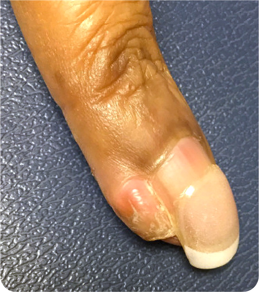
A 53-year-old patient presented with a painless lesion on the left third digit that had been slowly growing for about 20 years. The lesion arose after the patient removed an ingrown nail from the lateral nail bed. The cosmetic appearance of the nodule was bothersome to the patient, but it was otherwise asymptomatic. In 2008, the lesion was removed using shave excision, but it recurred and continued to grow slowly.
Physical examination revealed a hyperkeratotic, flesh-colored nodule measuring approximately 1 × 1 cm and located directly adjacent to the lateral nail bed of the left third digit (Figure 1). Plain radiographs of the finger revealed no cortical disruption, periosteal reaction, or suspicious lytic or blastic lesions. The patient had no systemic symptoms, unexplained weight loss, or other notable cutaneous lesions.
Question
Based on the patient's history and physical examination findings, which one of the following is the most likely diagnosis?
A. Acquired digital fibrokeratoma.
B. Dermatofibrosarcoma protuberans.
C. Glomus tumor.
D. Osteochondroma.
E. Superficial acral fibromyxoma.
Discussion
The answer is E: superficial acral fibromyxoma, a rare soft-tissue neoplasm. This patient has the typical presentation of a benign, slow-growing, firm, pink to flesh-colored nodule with a predilection for the periungual or subungual region of the digits or toes. The mean age at diagnosis is 50 years, and the male to female ratio is 1.3 to 1.1 The average tumor size at diagnosis is 1.7 cm (range of 0.5 cm to 5 cm).1,2 In one study, 41% of patients presented with a painful mass.1 Histologically, these tumors are well-circumscribed and comprised of short spindle cells in a myxoid stroma. The recurrence rate exceeds 20%.3 Complete surgical excision with clear margins is recommended.3
Acquired digital fibrokeratomas are benign, typically asymptomatic growths. They tend to be smaller, well-defined, hyperkeratotic, flesh-colored papules with a narrow stalk.4 They are characterized by a collarette of epidermis surrounding the stalk and histologically do not have a myxoid component.4,5
A glomus tumor is a rare, benign tumor arising from glomus bodies, which are dermal components involved in thermoregulation. The tumor most often presents as a solitary, blanchable, red-blue subungual or acral papule or nodule. It is typically painful, often in response to cold or pressure.
Osteochondromas, the most common benign bony tumors, usually present as painless growths in the long bones of children or adolescents. Imaging typically reveals a sessile or pedunculated mass arising from the metaphysis of the femur, tibia, or humerus, although they can occur on the hands and feet.
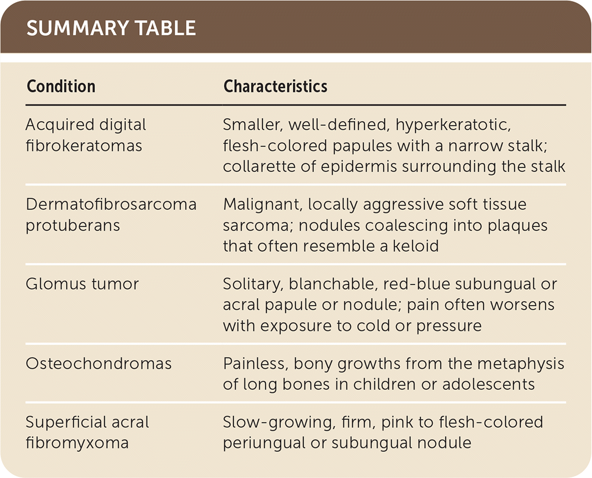
| Condition | Characteristics |
|---|---|
| Acquired digital fibrokeratomas | Smaller, well-defined, hyperkeratotic, flesh-colored papules with a narrow stalk; collarette of epidermis surrounding the stalk |
| Dermatofibrosarcoma protuberans | Malignant, locally aggressive soft tissue sarcoma; nodules coalescing into plaques that often resemble a keloid |
| Glomus tumor | Solitary, blanchable, red-blue subungual or acral papule or nodule; pain often worsens with exposure to cold or pressure |
| Osteochondromas | Painless, bony growths from the metaphysis of long bones in children or adolescents |
| Superficial acral fibromyxoma | Slow-growing, firm, pink to flesh-colored periungual or subungual nodule |
